


Frequently Bought Together
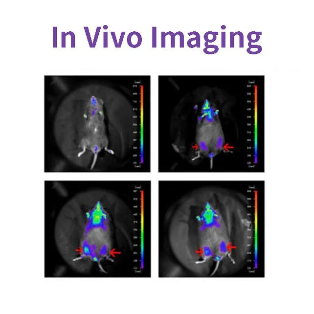
D-luciferin เป็นสารตั้งต้นทั่วไปสำหรับ Luciferase และใช้กันอย่างแพร่หลายในเทคโนโลยีชีวภาพ โดยเฉพาะเทคโนโลยีการถ่ายภาพในร่างกาย กลไกการทำงานคือ luciferin (สารตั้งต้น) สามารถออกซิไดซ์เพื่อปล่อยแสงในสภาพที่มี ATP และ luciferase เมื่อฟลูออเรสซีนมีมากเกินไป จำนวนควอนตัมของแสงที่ผลิตได้จะเท่ากับความเข้มข้นของ luciferase ที่มีความสัมพันธ์ในเชิงบวก (ดูรูปด้านล่าง) พลาสมิดที่มียีนเข้ารหัส luciferase (Luc) จะถูกถ่ายโอนเข้าสู่เซลล์และนำเข้าสู่สัตว์ที่ศึกษา เช่น หนูและหนูตะเภา

D-luciferin ยังใช้กันทั่วไปในการศึกษาในหลอดทดลอง รวมถึงการวิเคราะห์ระดับลูซิเฟอเรสและ ATP การวิเคราะห์ยีนรีพอร์ตเตอร์ การจัดลำดับประสิทธิภาพสูง และการทดสอบการปนเปื้อนต่างๆ มีรูปแบบผลิตภัณฑ์สามแบบ ได้แก่ D-luciferin (กรดอิสระ) เกลือ D-luciferin (เกลือโซเดียมและเกลือโพแทสเซียม) ความแตกต่างหลักอยู่ที่ความสามารถในการละลาย: ความสามารถในการละลายน้ำของ D-luciferin และความสามารถในการละลายของระบบบัฟเฟอร์ D-luciferin ละลายได้น้อย ยกเว้นในเบสอ่อน เช่น สารละลาย NaOH และ KOH ที่มีความเข้มข้นต่ำ ละลายได้ในเมทานอลและ DMSO ส่วน DMSO สามารถละลายในน้ำหรือบัฟเฟอร์ได้ง่าย ใช้งานง่าย
คุณสมบัติ
- ไม่มีรังสี แทบไม่มีอันตรายต่อสิ่งมีชีวิต
- การเรืองแสงชีวภาพ ไม่ต้องมีแหล่งกำเนิดแสงกระตุ้น
- ละเอียดอ่อนขนาดนี้ คุณสามารถตรวจพบได้ในเซลล์หลายร้อยเซลล์
- การเจาะที่ดี โดยยังสามารถตรวจจับความลึกของเนื้อเยื่อ 3-4 ซม. ได้
- อัตราส่วนสัญญาณต่อสัญญาณรบกวนสูง สัญญาณเรืองแสงที่แข็งแกร่ง และป้องกันสัญญาณรบกวนได้ดี
แอปพลิเคชั่น
- ในการทดลองการเกิดเนื้องอกในหนูเปลือย พบว่าการเติบโตของเนื้องอกเกิดขึ้นโดยไม่เกิดการบุกรุกแบบเรียลไทม์ โดยไม่ได้วัดการตัดเนื้องอก
- เพื่อทดสอบผลของการบริหารต่อการเจริญเติบโตของเนื้องอกหรือการแพร่กระจาย สารตั้งต้นฟลูออเรสซีนสามารถถูกกำจัดออกได้หมดภายใน 3 ชั่วโมง โดยไม่รบกวนการทดลองยา
- ตรวจพบตำแหน่งและการกระจายตัวของเซลล์แปลกปลอมในสัตว์
- ยีนเป้าหมายหรือโปรโมเตอร์ของยีนเป้าหมายจะถูกเชื่อมเข้ากับยีนลูซิเฟอเรสเพื่อตรวจจับการเปลี่ยนแปลงในการแสดงออกของยีนในระหว่างการรักษาด้วยยาหรือการดำเนินของโรค
- การติดตามการปลูกถ่าย การอยู่รอด และการแพร่กระจายของเซลล์ต้นกำเนิด ติดตามการกระจายและการอพยพของเซลล์ต้นกำเนิดในร่างกาย
ข้อมูลจำเพาะ
| คำพ้องความหมายภาษาอังกฤษ | (S)-4,5-Dihydro-2-(6-hydroxy-2-benzothiazolyl)-4-thiazolecarboxylic acid เกลือโพแทสเซียม; D-Luciferin หิ่งห้อย เกลือโพแทสเซียม |
| หมายเลข CAS | 115144-35-9 |
| สูตร | C11H7N2O3S2K |
| น้ำหนักโมเลกุล | 318.42 กรัม/โมล |
| รูปร่าง | ผงสีเหลืองอ่อน |
| ความสามารถในการละลาย | ละลายน้ำได้(60 มก./มล.) |
ส่วนประกอบ
| ส่วนประกอบ หมายเลข | ชื่อ | 40902ES01 | 40902ES02 | 40902ES03 | 40902ES08 |
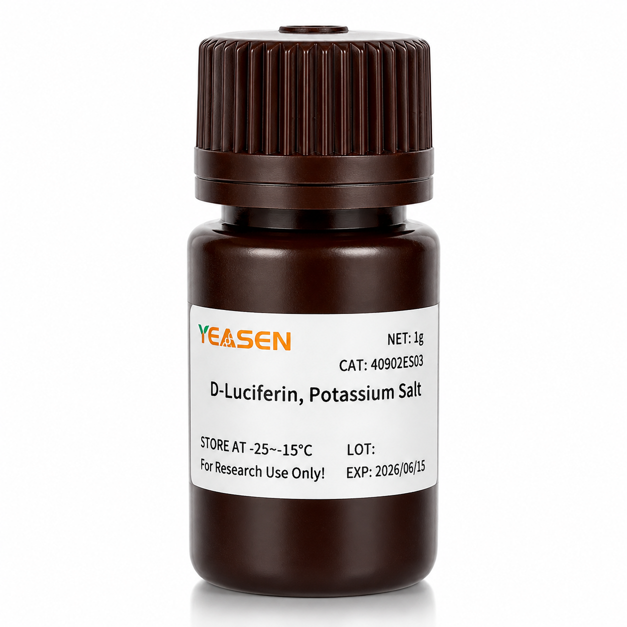
| 40902 | ดี-ลูซิเฟอริน เกลือโพแทสเซียม | 100 มก. | 500 มก. | 1กรัม | 5กรัม |
ตัวเลข

อ้างอิงจาก "A Novel Allosteric Inhibitor of Phosphoglycerate Mutase 1 Suppresses Growth and Metastasis of Non-Small-Cell Lung Cancer, Cell Metab.2019;30(6):1107-1119
การขนส่งและการเก็บรักษา
ผลิตภัณฑ์นี้จัดส่งพร้อมกับน้ำแข็งแห้งและสามารถจัดเก็บได้ที่อุณหภูมิ -15℃ ~ -25℃ สำหรับ 1 ปี.
[1] Gu Y, Wang Y, He L และคณะ Circular RNA circIPO11 drives self-renewal of liver cancer initiating cells via Hedgehog signaling. Mol Cancer. 2021;20(1):132. เผยแพร่เมื่อวันที่ 14 ตุลาคม 2021 doi:10.1186/s12943-021-01435-2(IF:27.401)
[2] Huang K, Liang Q, Zhou Y และคณะ สารยับยั้งอัลโลสเตอริกชนิดใหม่ของฟอสโฟกลีเซอเรตมิวเทส 1 ยับยั้งการเติบโตและการแพร่กระจายของมะเร็งปอดชนิดไม่ใช่เซลล์เล็ก [การแก้ไขที่เผยแพร่ปรากฏใน Cell Metab. 2021 ม.ค. 5;33(1):223] Cell Metab. 2019;30(6):1107-1119.e8 doi:10.1016/j.cmet.2019.09.014(IF:22.415)
[3] Dong X, Cheng R, Zhu S และคณะ นาโนไวต่อรังสี WO<sub>2.9</sub>-WSe<sub>2</sub> ที่มีโครงสร้างแบบเฮเทอโรจันก์ชั่นช่วยเพิ่มการทำลายเนื้องอกในบริเวณนั้นและภูมิคุ้มกันบำบัดด้วยการปิดกั้นจุดตรวจเมื่อได้รับรังสีในปริมาณต่ำ ACS Nano. 2020;14(5):5400-5416. doi:10.1021/acsnano.9b08962(IF:14.588)
[4] Zheng DW, Gao F, Cheng Q และคณะ ระบบนาโนที่ใช้วัคซีนเพื่อเริ่มต้นภูมิคุ้มกันโดยกำเนิดและปรับปรุงภูมิคุ้มกันบำบัดเนื้องอก Nat Commun. 2020;11(1):1985. เผยแพร่เมื่อวันที่ 24 เมษายน 2020 doi:10.1038/s41467-020-15927-0(IF:12.121)
[5] Hu J, Su H, Cao H และคณะ AUXIN RESPONSE FACTOR7 ผสานการส่งสัญญาณของจิบเบอเรลลินและออกซินผ่านปฏิสัมพันธ์ระหว่างโปรตีน DELLA และ AUX/IAA เพื่อควบคุมกิจกรรมของแคมเบียมในต้นป็อปลาร์ Plant Cell. 2022;34(7):2688-2707. doi:10.1093/plcell/koac107(IF:11.277)
[6] Qi P, Huang M, Hu X และคณะ เอฟเฟคเตอร์ของ Ralstonia solanacearum กำหนดเป้าหมายปัจจัยการถอดรหัส TGA เพื่อล้มล้างการส่งสัญญาณของกรดซาลิไซลิก เซลล์พืช 2022;34(5):1666-1683. ดอย:10.1093/plcell/koac015(IF:11.277)
[7] Huang X, Qiu M, Wang T และคณะ นาโนยาเอนกประสงค์ที่ปราศจากพาหะสำหรับการบำบัดมะเร็งรังไข่ที่แพร่กระจายทางช่องท้อง J Nanobiotechnology 2022;20(1):93 เผยแพร่เมื่อวันที่ 22 กุมภาพันธ์ 2022 doi:10.1186/s12951-022-01300-4(IF:10.435)
[8] Tang Y, Lin S, Yin S และคณะ การเกิดฟองก๊าซในแหล่งกำเนิดโดยอาศัยการย่อยสลายของอนุภาคแมกนีเซียม: แนวทางใหม่ในการผลิตไฮโดรเจลที่มีรูพรุนขนาดใหญ่ที่ฉีดได้ Biomaterials 2020;232:119727 doi:10.1016/j.biomaterials.2019.119727(IF:10.273)
[9] Zhang X, Zheng S, Hu C และคณะ lncRNA UPK1A-AS1 ที่เหนี่ยวนำโดยไฟโบรบลาสต์ที่เกี่ยวข้องกับมะเร็งช่วยให้เกิดความต้านทานแพลตตินัมในมะเร็งตับอ่อนผ่านการซ่อมแซมการแตกหักของสายคู่ที่มีประสิทธิภาพ Oncogene 2022;41(16):2372-2389 doi:10.1038/s41388-022-02253-6(IF:9.867)
[10] Qiao K, Liu Y, Xu Z และคณะ การเมทิลเลชัน RNA m6A ส่งเสริมการก่อตัวของการเลียนแบบหลอดเลือดในมะเร็งเซลล์ตับผ่านทาง Hippo pathway การสร้างหลอดเลือดใหม่ 2021;24(1):83-96 doi:10.1007/s10456-020-09744-8(IF:9.780)
[11] Wu Q, Kuang K, Lyu M และคณะ การปิดใช้งานอัลโลสเตอริกของ PIF และ EIN3 โดยไมโครโปรตีนในการควบคุมแสงของการพัฒนาของพืช Proc Natl Acad Sci US A. 2020;117(31):18858-18868 doi:10.1073/pnas.2002313117(IF:9.412)
[12] Meng DF, Sun R, Liu GY และคณะ S100A14 ยับยั้งการแพร่กระจายของมะเร็งโพรงหลังจมูกด้วยการยับยั้งการส่งสัญญาณ NF-kB ผ่านการย่อยสลายของ IRAK1 Oncogene 2020;39(30):5307-5322doi:10.1038/s41388-020-1363-8(แก้ไข:7.971)
[13] Gao Y, Wang J, Han H และคณะ วัสดุปลูกถ่ายโพลีคาโปรแลกโทนที่มีอนุภาคนาโนเพื่อต่อสู้กับการเกิดซ้ำของมะเร็งเต้านมหลังการผ่าตัด Nanoscale 2021;13(34):14417-14425 เผยแพร่เมื่อวันที่ 2 กันยายน 2021 doi:10.1039/d1nr04125h(IF:7.790)
[14] Jiang W, Li T, Guo J และคณะ เซลล์ CAR-T ที่มี c-Met/PD-L1 เฉพาะเจาะจงมีผลการรักษาที่ดีขึ้นต่อมะเร็งตับ Front Oncol 2021;11:546586 เผยแพร่เมื่อวันที่ 10 มีนาคม 2021 doi:10.3389/fonc.2021.546586(IF:6.244)
[15] Zhou Y , Zhou C , Zou Y , et al. ไมเซลล์ผสมคอนจูเกตยาที่ไวต่อค่า pH หลายชนิดสำหรับการนำส่งโดโซรูบิซินและเคอร์คูมินร่วมกันอย่างมีประสิทธิภาพเพื่อยับยั้งการแพร่กระจายของเนื้องอกอย่างมีประสิทธิภาพ Biomater Sci. 2020;8(18):5029-5046. doi:10.1039/d0bm00840k(IF:6.183)
[16] Zhao X, Liu X, Zhang P และคณะ ไฮโดรเจลเปปไทด์ฉีดเป็นไตรปโตไลด์ดีโปสำหรับฉีดเข้าช่องท้องเพื่อรักษามะเร็งตับในเซลล์ต้นกำเนิด Acta Pharm Sin B. 2019;9(5):1050-1060. doi:10.1016/j.apsb.2019.06.001(IF:5.808)
[17] Li M, Wang J, Yu Y และคณะ การกำหนดลักษณะของเซลล์ต้นกำเนิดของเนื้อเยื่อเกี่ยวพันที่ได้จากโรคกระดูกตายที่เกี่ยวข้องกับบิสฟอสโฟเนตของเหงือกของผู้ป่วยขากรรไกร Stem Cell Rev Rep. 2022;18(1):378-394 doi:10.1007/s12015-021-10241-8(IF:5.739)
[18] Zhang X, Li Y, Ji J และคณะ Gadd45g ริเริ่มการแบ่งตัวของเซลล์ต้นกำเนิดตัวอ่อนและยับยั้งการก่อมะเร็งเซลล์เต้านม Cell Death Discov. 2021;7(1):271. เผยแพร่เมื่อวันที่ 2 ตุลาคม 2021 doi:10.1038/s41420-021-00667-x(IF:5.241)
[19] Wu D, Lv J, Zhao R และคณะ PSCA เป็นเป้าหมายของเซลล์ T ของตัวรับแอนติเจนไคเมอริกในมะเร็งกระเพาะอาหาร Biomark Res. 2020;8:3. เผยแพร่เมื่อวันที่ 28 มกราคม 2020 doi:10.1186/s40364-020-0183-x(IF:4.866)
[20] Qiao K, Chen C, Liu H, Qin Y, Liu H. Pinin ชักนำการเปลี่ยนผ่านจากเยื่อบุผิวไปเป็นเนื้อเยื่อเกี่ยวพันในมะเร็งตับโดยควบคุมการดัดแปลง m6A J Oncol. 2021;2021:7529164 เผยแพร่เมื่อวันที่ 7 ธันวาคม 2021 doi:10.1155/2021/7529164(IF:4.375)
[21] Li K, Liu T, Chen J, Ni H, Li W. Survivin ในเอ็กโซโซมที่ได้มาจากมะเร็งเต้านมกระตุ้นไฟโบรบลาสต์โดยเพิ่มการทำงานของ SOD1 ซึ่งการตอบสนองของ SOD1 จะส่งเสริมการแพร่กระจายและแพร่กระจายของมะเร็ง J Biol Chem. 2020;295(40):13737-13752. doi:10.1074/jbc.RA120.013805(IF:4.238)
[22] Kong Y, Feng Z, Chen A และคณะ ฟลาโวนอยด์กาแลงจินจากธรรมชาติกระตุ้นการเกิดอะพอพโทซิส ไพโรพโทซิส และออโตฟาจีในกลีโอบลาสโตมา Front Oncol 2019;9:942 เผยแพร่เมื่อวันที่ 27 กันยายน 2019 doi:10.3389/fonc.2019.00942(IF:4.137)
[23] Kong Y, Feng Z, Chen A และคณะ ฟลาโวนอยด์กาแลงจินจากธรรมชาติกระตุ้นการเกิดอะพอพโทซิส ไพโรพโทซิส และออโตฟาจีในกลีโอบลาสโตมา Front Oncol 2019;9:942 เผยแพร่เมื่อวันที่ 27 กันยายน 2019 doi:10.3389/fonc.2019.00942(IF:4.137)
[24] Hu J, Wang Y, Yuan Y. สารยับยั้งการทำงานของ APE1 redox ยับยั้งการจำลองแบบของγ-herpesvirus ได้อย่างมีประสิทธิภาพทั้งในหลอดทดลองและในร่างกาย Antiviral Res. 2021;185:104985. doi:10.1016/j.antiviral.2020.104985(IF:4.101)
[25] Li Q, Wei D, Feng F และคณะ กรดซาลิกที่เชื่อมโยงกับ α2,6 ทำหน้าที่เป็นตัวรับที่มีความสัมพันธ์สูงสำหรับการบำบัดด้วยไวรัสที่ทำลายเซลล์มะเร็งด้วยไวรัสโรคนิวคาสเซิล J Cancer Res Clin Oncol. 2017;143(11):2171-2181. doi:10.1007/s00432-017-2470-y(IF:3.503)
[26] Han B, Jiang P, Liu W และคณะ บทบาทของ Daucosterol Linoleate ต่อมะเร็งเต้านม: การศึกษาเกี่ยวกับอะพอพโทซิสและการแพร่กระจาย J Agric Food Chem. 2018;66(24):6031-6041 doi:10.1021/acs.jafc.8b01387(IF:3.412)
[27] Hu H, Zhang Z, Wang R และคณะ เซลล์ BGC823 ที่มีการแสดงออกที่เสถียรของ iRFP720 ยังคงคุณสมบัติหลักไว้ด้วยความสามารถในการสร้างภาพฟลูออเรสเซนต์ที่มีแนวโน้มดี DNA Cell Biol. 2020;39(5):900-908. doi:10.1089/dna.2019.5057(IF:3.314)
(28) Ni XR, Zhao YY, Cai HP และอื่น ๆการถ่ายภาพด้วยแสงที่กำหนดเป้าหมายตัวรับทรานสเฟอร์ริน 1 เพื่อระบุขอบของก้อนเนื้อในสมองในแบบจำลองเมาส์ J Neurooncol 2020;148(2):245-258 doi:10.1007/s11060-020-03527-3(IF:3.267)
[29] Feng H, Tang J, Zhang P, Miao Y, Wu T, Cheng Z. สเตอริโอไอโซเมอร์ 18,19-seco-ursane ที่ต่อต้านการเกิดไขมันและซาโปนินชนิดโอลีเอนจากใบ Ilex cornuta Phytochemistry. 2020;175:112363. doi:10.1016/j.phytochem.2020.112363(IF:3.044)
[30] Zhao Y, Li T, Tian S และคณะ การยับยั้ง MYC-Amplified Group 3 Medulloblastoma ได้อย่างมีประสิทธิภาพผ่านการกำหนดเป้าหมาย EIF4A1 Cancer Manag Res. 2020;12:12473-12485 เผยแพร่เมื่อวันที่ 3 ธันวาคม 2020 doi:10.2147/CMAR.S278844(IF:2.886)
Storage
FAQ
1) Solubility: The salt form is easily soluble in water. The solubility of potassium salt is 60mg/ml, and that of sodium salt is 100mg/ml. Free acid is not easily soluble in water. It can be dissolved by a weak alkaline solution of sodium bicarbonate, and its solubility in methanol is 10mg/ml, and in DMSO is 50mg/ml.
2) Toxicity: The salt form is more convenient to use during the process, especially in in vivo imaging experiments, as it can dissolve in water and the reaction toxicity will be smaller (Fluorescein is a low-molecular-weight organic compound composed of phenylthiazole and thiazole carboxyl group, with low toxicity).
3) Usage effect: There is no significant difference. In in vivo experimental studies, the usage rate of potassium salt is higher.
2. Native coelenterazine is an action substrate for various luciferases such as sea kidney luciferase (Rluc) and Gaussia luciferase (Gluc).
1. Instruments with a biochemiluminescence detection module. The light produced by luciferin can be detected by a photometer or a scintillation counter. Common in vivo imaging instruments: such as the IVIS® Lumina small animal in vivo imaging system, the In-Vivo Xtreme multi-mode small animal in vivo imaging instrument from Bruker, Germany.
2. Multi-functional microplate readers: It is necessary to confirm with the instrument manufacturer whether it is a specific biochemiluminescence detection module (note: cannot use a fluorescence microscope).
2. The cells themselves also contain a certain amount of ATP and magnesium, which can promote the reaction. In most cases, the ATP and magnesium contained in the cells are sufficient for the reaction of D-fluorescein salt, but the content varies depending on the type of cells. You can first try using a culture medium without additional ATP and magnesium, and conduct the experiment normally to see the results.
1) Injection method: It can be done through intraperitoneal injection or tail vein injection.
2) Injection volume: The scientific approach is to evaluate the injection dose based on the kinetic curve. The initial attempted injection dose is: 150mg substrate/kg of mouse body weight. Therefore, the purchase quantity can be calculated according to the above method: If there are 10 mice, 22 - 25g, then 33 - 37.5mg of substrate is required.
Citations
Documents
| Brochure |
|---|
| In_vivo_imaging_Leaving_Tumor_Cells_Nowhere_to_Hide.pdf |


คุณอาจชอบ
การสอบถาม
คำถามที่พบบ่อย
ผลิตภัณฑ์นี้มีวัตถุประสงค์เพื่อการวิจัยเท่านั้น และไม่ได้มีวัตถุประสงค์เพื่อใช้ในการรักษาหรือวินิจฉัยโรคในมนุษย์หรือสัตว์ ผลิตภัณฑ์และเนื้อหาได้รับการคุ้มครองโดยสิทธิบัตร เครื่องหมายการค้า และลิขสิทธิ์ที่เป็นของ
แอปพลิเคชั่นบางตัวอาจต้องใช้สิทธิ์ในทรัพย์สินทางปัญญาของบุคคลที่สามเพิ่มเติม