


Frequently Bought Together
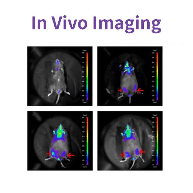
D-luciferin är ett vanligt substrat för Luciferas och används i stor utsträckning inom bioteknik, särskilt in vivo-avbildningsteknik. Verkningsmekanismen är att luciferin (substrat) kan oxideras för att avge ljus i närvaro av ATP och luciferas. När fluorescein är i överskott, är kvantantalet ljus som produceras lika med Luciferas-koncentrationerna var positivt korrelerade (se figur nedan). Plasmider som bär på luciferaskodande gen (Luc) transfekterades in i celler och introducerades i studiedjur såsom råttor och möss.

D-luciferin används också ofta i in vitro-studier, inklusive luciferas- och ATP-nivåanalys; Reportergenanalys; Sekvensering med hög genomströmning och olika kontamineringstester. Det finns tre produktformer: D-luciferin (fri syra), D-luciferinsalt (natriumsalt och kaliumsalt). Huvudskillnaden ligger i lösligheten: vattenlösligheten hos den förra och lösligheten hos buffertsystemet. De är svagt lösliga förutom i svaga baser som låga koncentrationer av NaOH- och KOH-lösningar. Löslig i metanol och DMSO; Den senare kan lätt lösas i vatten eller buffert, lätt att använda.
Drag
- Ingen strålning, nästan ofarlig för levande organismer.
- Bioluminescens, ingen excitationsljuskälla.
- Så känslig att du kan upptäcka den i några hundra celler.
- Bra penetration, 3-4 cm vävnadsdjup kan fortfarande detekteras.
- Högt signal-brusförhållande, stark fluorescenssignal och bra anti-interferens.
Ansökningar
- I tumörbildningsexperimentet i nakna möss observerades tumörtillväxten utan invasion i realtid, utan tumöravdrivningsmätning.
- För att testa effekten av administreringen på tumörtillväxt eller metastasering kan fluoresceinsubstratet elimineras fullständigt inom 3 timmar, utan störning av läkemedelsexperimentet.
- Lokaliseringen och distributionen av främmande celler i djur detekterades.
- Målgenen eller promotorn för målgenen är sammansmält med luciferasgenen för att detektera förändringar i genuttryck under läkemedelsbehandling eller sjukdomsprogression.
- Övervakning av stamcellstransplantation, överlevnad och spridning; Spåra distributionen och migrationen av stamceller in vivo.
Specifikationer
| Engelsk synonym | (S)-4,5-dihydro-2-(6-hydroxi-2-bensotiazolyl)-4-tiazolkarboxylsyra kaliumsalt; D-Luciferin eldfluga, kaliumsalt |
| CAS NR. | 115144-35-9 |
| Formel | C11H7N2O3S2K |
| Molekylvikt | 318,42 g/mol |
| Utseende | Ljusgult pulver |
| Löslighet | Lösligt i vatten (60 mg/ml) |
Komponenter
| Komponentnr. | Namn | 40902ES01 | 40902ES02 | 40902ES03 | 40902ES08 |
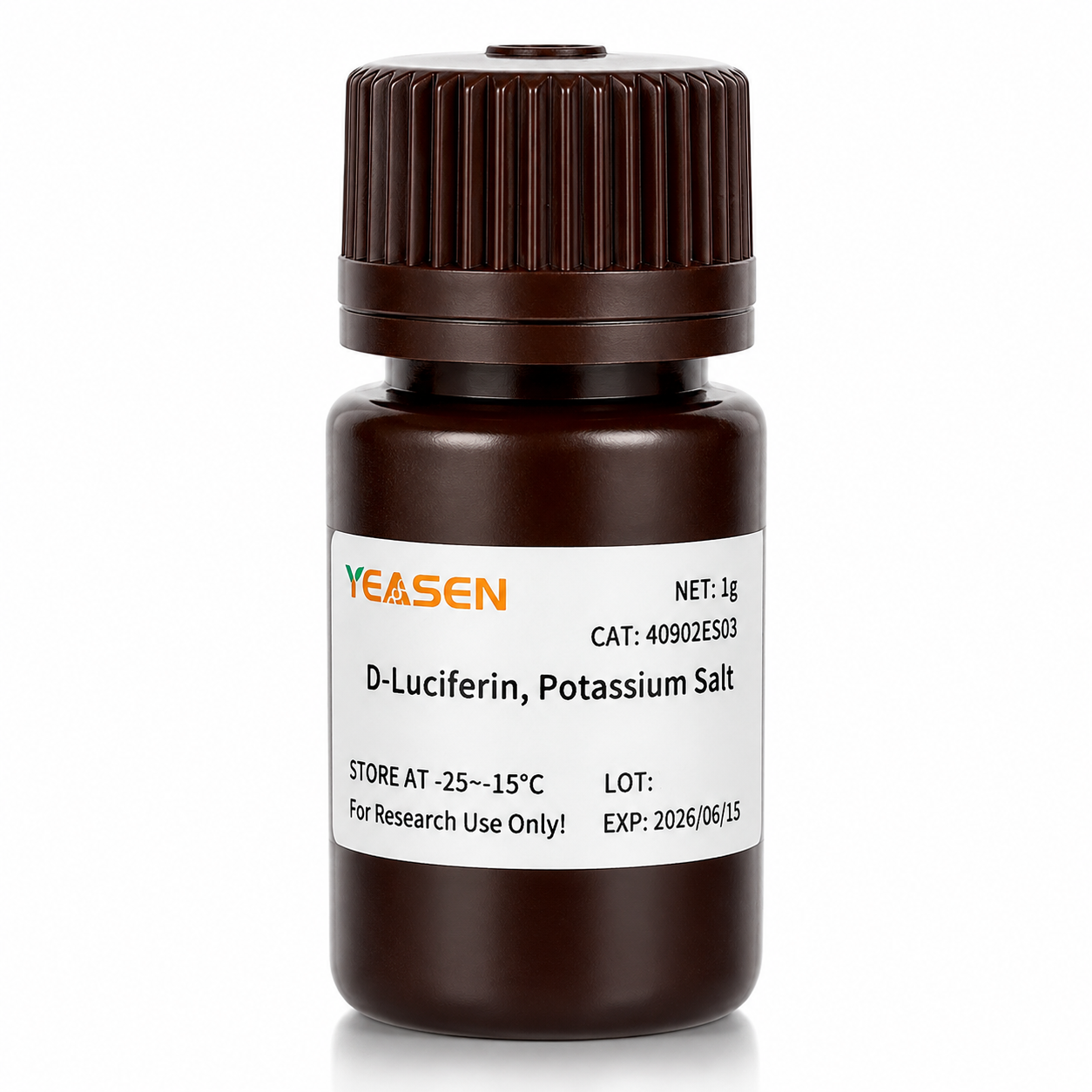
| 40902 | D-Luciferin, Kaliumsalt | 100 mg | 500 mg | 1g | 5g |
Siffror

Citerad från "A Novel Allosteric Inhibitor of Phosphoglycerate Mutase 1 undertrycker tillväxt och metastasering av icke-småcellig lungcancer, Cell Metab.2019;30(6):1107-1119
Frakt och förvaring
Produkten levereras med torris och kan förvaras vid -15℃ ~ -25℃ för 1 år.
[1] Gu Y, Wang Y, He L, et al. Cirkulärt RNA circIPO11 driver självförnyelse av levercancerinitierande celler via Hedgehog-signalering. Mol Cancer. 2021;20(1):132. Publicerad 2021 14 oktober. doi:10.1186/s12943-021-01435-2(IF:27.401)
[2] Huang K, Liang Q, Zhou Y, et al. En ny allosterisk hämmare av fosfoglyceratmutas 1 undertrycker tillväxt och metastasering av icke-småcellig lungcancer [publicerad korrigering visas i Cell Metab. 2021 jan 5;33(1):223]. Cell Metab. 2019;30(6):1107-1119.e8. doi:10.1016/j.cmet.2019.09.014(IF:22.415)
[3] Dong X, Cheng R, Zhu S, et al. En Heterojunction Structured WO<sub>2.9</sub>-WSe<sub>2</sub> Nanoradiosensibilisator ökar lokal tumörablation och kontrollpunktsblockad immunterapi vid låg stråldos. ACS Nano. 2020;14(5):5400-5416. doi:10.1021/acsnano.9b08962(IF:14.588)
[4] Zheng DW, Gao F, Cheng Q, et al. Ett vaccinbaserat nanosystem för att initiera medfödd immunitet och förbättra tumörimmunterapi. Nat Commun. 2020;11(1):1985. Publicerad 2020 apr 24. doi:10.1038/s41467-020-15927-0(IF:12.121)
[5] Hu J, Su H, Cao H, et al. AUXIN RESPONSE FACTOR7 integrerar gibberellin- och auxinsignalering via interaktioner mellan DELLA- och AUX/IAA-proteiner för att reglera kambial aktivitet i poppel. Växtcell. 2022;34(7):2688-2707. doi:10.1093/plcell/koac107(IF:11.277)
[6] Qi P, Huang M, Hu X, et al. En Ralstonia solanacearum-effektor riktar sig mot TGA-transkriptionsfaktorer för att undergräva salicylsyrasignalering. Växtcell. 2022;34(5):1666-1683. doi:10.1093/plcell/koac015(IF:11.277)
[7] Huang X, Qiu M, Wang T, et al. Bärarfri multifunktionell nanomedicin för intraperitoneal spridd äggstockscancerterapi. J Nanobioteknologi. 2022;20(1):93. Publicerad 2022 22 februari doi:10.1186/s12951-022-01300-4(IF:10.435)
[8] Tang Y, Lin S, Yin S, et al. In situ gasskumning baserad på magnesiumpartikelnedbrytning: En ny metod för att tillverka injicerbara makroporösa hydrogeler. Biomaterial. 2020;232:119727. doi:10.1016/j.biomaterials.2019.119727(IF:10.273)
[9] Zhang X, Zheng S, Hu C, et al. Cancerassocierad fibroblastinducerad lncRNA UPK1A-AS1 ger platinaresistens vid pankreascancer via effektiv dubbelsträngsbrottsreparation. Onkogen. 2022;41(16):2372-2389. doi:10.1038/s41388-022-02253-6(IF:9.867)
[10] Qiao K, Liu Y, Xu Z, et al. RNA m6A-metylering främjar bildandet av vaskulogen mimik i hepatocellulärt karcinom via Hippo-vägen. Angiogenes. 2021;24(1):83-96. doi:10.1007/s10456-020-09744-8(IF:9.780)
[11] Wu Q, Kuang K, Lyu M, et al. Allosterisk deaktivering av PIF och EIN3 av mikroproteiner i ljuskontroll av växtutveckling. Proc Natl Acad Sci US A. 2020;117(31):18858-18868. doi:10.1073/pnas.2002313117(IF:9.412)
[12] Meng DF, Sun R, Liu GY, et al. S100A14 undertrycker metastasering av nasofarynxkarcinom genom hämning av NF-kB-signalering genom nedbrytning av IRAK1. Onkogen. 2020;39(30):5307-5322.doi:10.1038/s41388-020-1363-8(IF:7.971)
[13] Gao Y, Wang J, Han H, et al. Ett nanopartikelinnehållande polykaprolaktonimplantat för att bekämpa återfall av bröstcancer efter resektion. Nanoskala. 2021;13(34):14417-14425. Publicerad 2021 september 2. doi:10.1039/d1nr04125h(IF:7.790)
[14] Jiang W, Li T, Guo J, et al. Bispecifika c-Met/PD-L1 CAR-T-celler har förbättrade terapeutiska effekter på hepatocellulärt karcinom. Front Oncol. 2021;11:546586. Publicerad 2021 10 mars. doi:10.3389/fonc.2021.546586(IF:6.244)
[15] Zhou Y, Zhou C, Zou Y, et al. Multi pH-känsliga polymer-läkemedelskonjugat blandade miceller för effektiv samtidig leverans av doxorubicin och curcumin för att synergistiskt undertrycka tumörmetastaser. Biomater Sci. 2020;8(18):5029-5046. doi:10.1039/d0bm00840k(IF:6.183)
[16] Zhao X, Liu X, Zhang P, et al. Injicerbar peptidhydrogel som intraperitoneal triptoliddepå för behandling av ortotopiskt hepatocellulärt karcinom. Acta Pharm Sin B. 2019;9(5):1050-1060. doi:10.1016/j.apsb.2019.06.001(IF:5.808)
[17] Li M, Wang J, Yu Y, et al. Karakterisering av mesenkymala stamceller härledda från bisfosfonatrelaterad osteonekros i käkpatienternas gingiva. Stamcell Rev Rep. 2022;18(1):378-394. doi:10.1007/s12015-021-10241-8(IF:5.739)
[18] Zhang X, Li Y, Ji J, et al. Gadd45g initierar embryonal stamcellsdifferentiering och hämmar bröstcellscancer. Celldöd Discov. 2021;7(1):271. Publicerad 2 oktober 2021. doi:10.1038/s41420-021-00667-x(IF:5.241)
[19] Wu D, Lv J, Zhao R, et al. PSCA är ett mål för chimära antigenreceptor-T-celler i magcancer. Biomark Res. 2020;8:3. Publicerad 2020 28 januari. doi:10.1186/s40364-020-0183-x(IF:4.866)
[20] Qiao K, Chen C, Liu H, Qin Y, Liu H. Pinin inducerar epitel-till-mesenkymal övergång i hepatocellulärt karcinom genom att reglera m6A-modifiering. J Oncol. 2021;2021:7529164. Publicerad 2021 7 dec. doi:10.1155/2021/7529164(IF:4.375)
[21] Li K, Liu T, Chen J, Ni H, Li W. Survivin i bröstcancer-härledda exosomer aktiverar fibroblaster genom att uppreglera SOD1, vars feedback främjar cancerproliferation och metastasering. J Biol Chem. 2020;295(40):13737-13752. doi:10.1074/jbc.RA120.013805(IF:4.238)
[22] Kong Y, Feng Z, Chen A, et al. Den naturliga flavonoiden Galangin framkallar apoptos, pyroptos och autofagi i glioblastom. Front Oncol. 2019;9:942. Publicerad 2019 27 september doi:10.3389/fonc.2019.00942(IF:4.137)
[23] Kong Y, Feng Z, Chen A, et al. Den naturliga flavonoiden Galangin framkallar apoptos, pyroptos och autofagi i glioblastom. Front Oncol. 2019;9:942. Publicerad 2019 27 september doi:10.3389/fonc.2019.00942(IF:4.137)
[24] Hu J, Wang Y, Yuan Y. Hämmare av APE1 redoxfunktion hämmar effektivt γ-herpesvirusreplikation in vitro och in vivo. Antiviral Res. 2021;185:104985. doi:10.1016/j.antiviral.2020.104985(IF:4.101)
[25] Li Q, Wei D, Feng F, et al. α2,6-kopplad sialinsyra fungerar som en högaffinitetsreceptor för canceronkolytisk viroterapi med viruset Newcastlesjuka. J Cancer Res Clin Oncol. 2017;143(11):2171-2181. doi:10.1007/s00432-017-2470-y(IF:3.503)
[26] Han B, Jiang P, Liu W, et al. Roll av Daucosterol Linoleate på bröstcancer: Studier om apoptos och metastaser. J Agric Food Chem. 2018;66(24):6031-6041. doi:10.1021/acs.jafc.8b01387(IF:3.412)
[27] Hu H, Zhang Z, Wang R, et al. BGC823-cellinje med det stabila uttrycket av iRFP720 behåller sina primära egenskaper med lovande fluorescensavbildningsförmåga. DNA Cell Biol. 2020;39(5):900-908. doi:10.1089/dna.2019.5057(IF:3.314)
[28] Ni XR, Zhao YY, Cai HP, et al.Transferrinreceptor 1 riktad optisk avbildning för identifiering av gliommarginal i musmodeller. J Neuroncol. 2020;148(2):245-258. doi:10.1007/s11060-020-03527-3(IF:3.267)
[29] Feng H, Tang J, Zhang P, Miao Y, Wu T, Cheng Z. Anti-adipogena 18,19-seco-ursane stereoisomerer och saponiner av oleantyp från Ilex cornuta-blad. Fytokemi. 2020;175:112363. doi:10.1016/j.phytochem.2020.112363(IF:3.044)
[30] Zhao Y, Li T, Tian S, et al. Effektiv hämning av MYC-förstärkt grupp 3-medulloblastom genom inriktning på EIF4A1. Cancer Manag Res. 2020;12:12473-12485. Publicerad 2020 3 dec. doi:10.2147/CMAR.S278844(IF:2.886)
Storage
FAQ
1) Solubility: The salt form is easily soluble in water. The solubility of potassium salt is 60mg/ml, and that of sodium salt is 100mg/ml. Free acid is not easily soluble in water. It can be dissolved by a weak alkaline solution of sodium bicarbonate, and its solubility in methanol is 10mg/ml, and in DMSO is 50mg/ml.
2) Toxicity: The salt form is more convenient to use during the process, especially in in vivo imaging experiments, as it can dissolve in water and the reaction toxicity will be smaller (Fluorescein is a low-molecular-weight organic compound composed of phenylthiazole and thiazole carboxyl group, with low toxicity).
3) Usage effect: There is no significant difference. In in vivo experimental studies, the usage rate of potassium salt is higher.
2. Native coelenterazine is an action substrate for various luciferases such as sea kidney luciferase (Rluc) and Gaussia luciferase (Gluc).
1. Instruments with a biochemiluminescence detection module. The light produced by luciferin can be detected by a photometer or a scintillation counter. Common in vivo imaging instruments: such as the IVIS® Lumina small animal in vivo imaging system, the In-Vivo Xtreme multi-mode small animal in vivo imaging instrument from Bruker, Germany.
2. Multi-functional microplate readers: It is necessary to confirm with the instrument manufacturer whether it is a specific biochemiluminescence detection module (note: cannot use a fluorescence microscope).
2. The cells themselves also contain a certain amount of ATP and magnesium, which can promote the reaction. In most cases, the ATP and magnesium contained in the cells are sufficient for the reaction of D-fluorescein salt, but the content varies depending on the type of cells. You can first try using a culture medium without additional ATP and magnesium, and conduct the experiment normally to see the results.
1) Injection method: It can be done through intraperitoneal injection or tail vein injection.
2) Injection volume: The scientific approach is to evaluate the injection dose based on the kinetic curve. The initial attempted injection dose is: 150mg substrate/kg of mouse body weight. Therefore, the purchase quantity can be calculated according to the above method: If there are 10 mice, 22 - 25g, then 33 - 37.5mg of substrate is required.
Citations
Documents
| Brochure |
|---|
| In_vivo_imaging_Leaving_Tumor_Cells_Nowhere_to_Hide.pdf |


Du kanske också gillar
Förfrågan
Vanliga frågor
Produkten är endast avsedd för forskningsändamål och är inte avsedd för terapeutisk eller diagnostisk användning hos människor eller djur. Produkter och innehåll skyddas av patent, varumärken och upphovsrätt som ägs av Yeasen Biotechnology. Varumärkessymboler anger ursprungsland, inte nödvändigtvis registrering i alla regioner.
Vissa applikationer kan kräva ytterligare immateriella rättigheter från tredje part.
Yeasen är dedikerad till etisk vetenskap, och anser att vår forskning bör behandla kritiska frågor samtidigt som den säkerställer säkerhet och etiska standarder.