


Frequently Bought Together
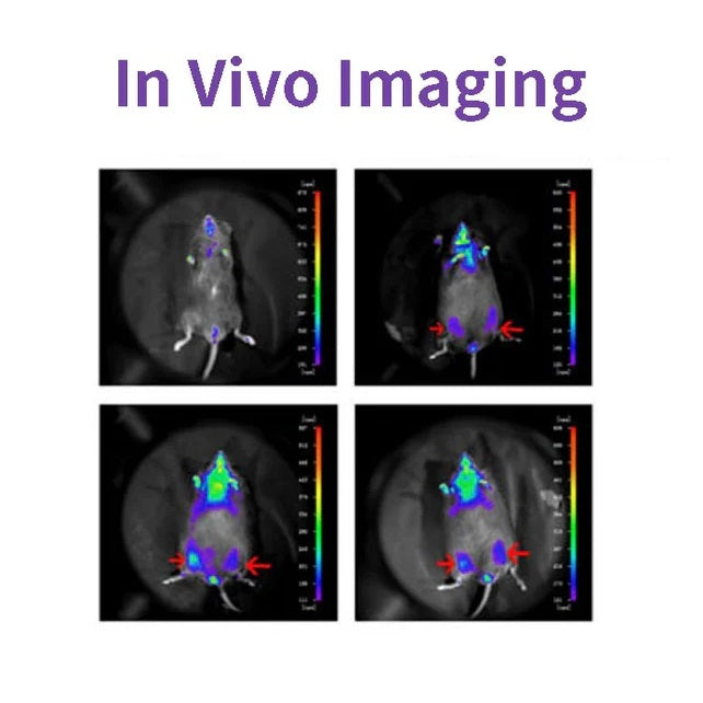
D-luciferine is een veelgebruikt substraat voor Luciferase en wordt veel gebruikt in de biotechnologie, met name in vivo imaging Technology. Het werkingsmechanisme is dat luciferine (substraat) kan worden geoxideerd om licht uit te zenden in aanwezigheid van ATP en luciferase. Wanneer fluoresceïne in overmaat aanwezig is, is het kwantumgetal van het geproduceerde licht gelijk aan Luciferaseconcentraties waren positief gecorreleerd (zie onderstaande afbeelding). Plasmiden die het luciferase-coderende gen (Luc) dragen, werden getransfecteerd in cellen en geïntroduceerd in proefdieren zoals ratten en muizen.

D-luciferine wordt ook vaak gebruikt in in vitro-onderzoeken, waaronder luciferase- en ATP-niveauanalyse; Reportergenanalyse; High-throughput sequencing en verschillende contaminatietesten. Er zijn drie productvormen: D-luciferine (vrij zuur), D-luciferinezout (natriumzout en kaliumzout). Het belangrijkste verschil ligt in de oplosbaarheid: wateroplosbaarheid van de eerste en oplosbaarheid van het buffersysteem. Ze zijn zwak oplosbaar, behalve in zwakke basen zoals lage concentraties NaOH- en KOH-oplossingen. Oplosbaar in methanol en DMSO; de laatste kan gemakkelijk worden opgelost in water of buffer, gemakkelijk te gebruiken.
Functies
- Geen straling, vrijwel ongevaarlijk voor levende organismen.
- Bioluminescentie, geen exciterende lichtbron.
- Zo gevoelig dat je het in een paar honderd cellen kunt detecteren.
- Goede penetratie, weefseldiepte van 3-4 cm is nog steeds te detecteren.
- Hoge signaal-ruisverhouding, sterk fluorescentiesignaal en goede anti-interferentie.
Toepassingen
- Bij het tumorigenese-experiment bij naakte muizen werd de tumorgroei in realtime waargenomen zonder invasie, zonder dat de tumor werd afgestoten.
- Om het effect van de toediening op de tumorgroei of metastasering te testen, kan het fluoresceïnesubstraat binnen 3 uur volledig worden verwijderd, zonder dat het medicijnexperiment wordt verstoord.
- De lokalisatie en distributie van vreemde cellen bij dieren werden gedetecteerd.
- Het doelgen of de promotor van het doelgen wordt gefuseerd met het luciferasegen om veranderingen in de genexpressie te detecteren tijdens een medicijnbehandeling of ziekteprogressie.
- Toezicht op stamceltransplantatie, overleving en proliferatie; Volg de distributie en migratie van stamcellen in vivo.
Specificaties
| Engels synoniem | (S)-4,5-Dihydro-2-(6-hydroxy-2-benzothiazolyl)-4-thiazoolcarbonzuur kaliumzout; D-Luciferin vuurvlieg, kaliumzout |
| CAS-NR. | 115144-35-9 |
| Formule | C11H7N2O3S2K |
| Moleculair gewicht | 318,42 gram/mol |
| Verschijning | Lichtgeel poeder |
| Oplosbaarheid | Oplosbaar in water (60 mg/ml) |
Componenten
| Componenten nr. | Naam | 40902ES01 | 40902ES02 | 40902ES03 | 40902ES08 |
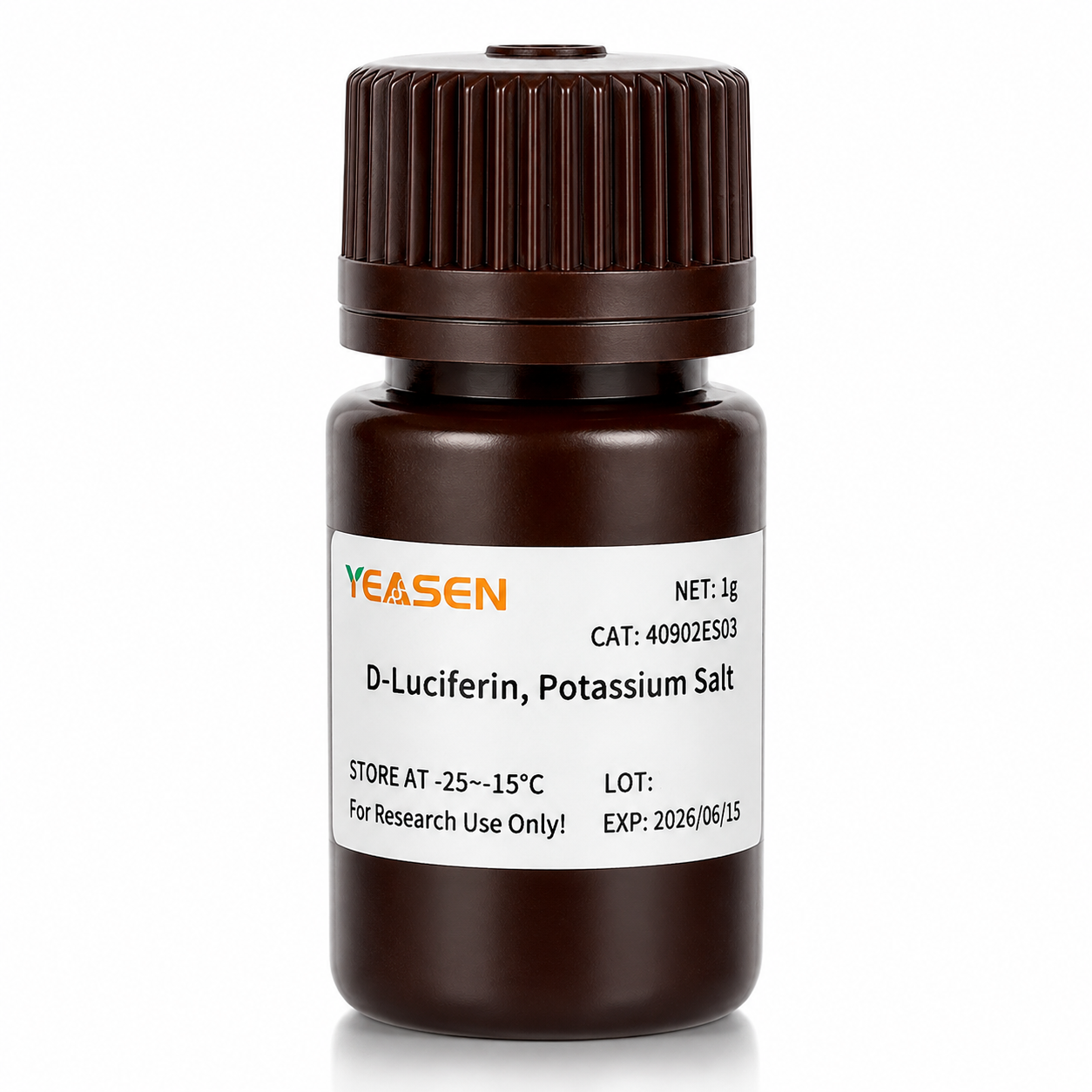
| 40902 | D-Luciferine, Kaliumzout | 100mg | 500mg | 1 gram | 5 gram |
Cijfers

Geciteerd uit "Een nieuwe allosterische remmer van fosfoglyceraatmutase 1 onderdrukt de groei en metastase van niet-kleincellige longkanker, celmetab.2019;30(6):1107-1119
Verzending en opslag
Het product wordt verzonden met droogijs en kan worden bewaard bij -15℃ ~ -25℃ voor 1 jaar.
[1] Gu Y, Wang Y, He L, et al. Circulair RNA circIPO11 drijft zelfvernieuwing van leverkanker-initiërende cellen aan via Hedgehog-signalering. Mol Cancer. 2021;20(1):132. Gepubliceerd 14 okt. 2021. doi:10.1186/s12943-021-01435-2(IF:27.401)
[2] Huang K, Liang Q, Zhou Y, et al. Een nieuwe allosterische remmer van fosfoglyceraatmutase 1 onderdrukt de groei en metastase van niet-kleincellige longkanker [gepubliceerde correctie verschijnt in Cell Metab. 2021 5 jan;33(1):223]. Cell Metab. 2019;30(6):1107-1119.e8. doi:10.1016/j.cmet.2019.09.014(IF:22.415)
[3] Dong X, Cheng R, Zhu S, et al. Een heterojunctie-gestructureerde WO<sub>2.9</sub>-WSe<sub>2</sub> nanoradiosensitizer verhoogt lokale tumorablatie en checkpointblokkade-immunotherapie bij lage stralingsdosis. ACS Nano. 2020;14(5):5400-5416. doi:10.1021/acsnano.9b08962(IF:14.588)
[4] Zheng DW, Gao F, Cheng Q, et al. Een op vaccins gebaseerd nanosysteem voor het initiëren van aangeboren immuniteit en het verbeteren van tumorimmunotherapie. Nat Commun. 2020;11(1):1985. Gepubliceerd 2020 Apr 24. doi:10.1038/s41467-020-15927-0(IF:12.121)
[5] Hu J, Su H, Cao H, et al. AUXIN RESPONSE FACTOR7 integreert gibberelline- en auxinesignalering via interacties tussen DELLA- en AUX/IAA-eiwitten om cambiale activiteit in populier te reguleren. Plant Cell. 2022;34(7):2688-2707. doi:10.1093/plcell/koac107(IF:11.277)
[6] Qi P, Huang M, Hu X, et al. Een Ralstonia solanacearum-effector richt zich op TGA-transcriptiefactoren om de salicylzuursignalering te ondermijnen. Plantencel. 2022;34(5):1666-1683. doi:10.1093/plcell/koac015(IF:11.277)
[7] Huang X, Qiu M, Wang T, et al. Dragervrije multifunctionele nanomedicijnen voor intraperitoneale gedissemineerde ovariumkankertherapie. J Nanobiotechnology. 2022;20(1):93. Gepubliceerd 22 feb. 2022. doi:10.1186/s12951-022-01300-4(IF:10.435)
[8] Tang Y, Lin S, Yin S, et al. In situ gasschuiming op basis van magnesiumdeeltjesafbraak: een nieuwe benadering om injecteerbare macroporeuze hydrogels te fabriceren. Biomaterials. 2020;232:119727. doi:10.1016/j.biomaterials.2019.119727(IF:10.273)
[9] Zhang X, Zheng S, Hu C, et al. Kanker-geassocieerde fibroblast-geïnduceerde lncRNA UPK1A-AS1 verleent platinaresistentie bij alvleesklierkanker via efficiënte reparatie van dubbelstrengsbreuken. Oncogene. 2022;41(16):2372-2389. doi:10.1038/s41388-022-02253-6(IF:9.867)
[10] Qiao K, Liu Y, Xu Z, et al. RNA m6A-methylering bevordert de vorming van vasculogene mimicry in hepatocellulair carcinoom via het Hippo-pad. Angiogenese. 2021;24(1):83-96. doi:10.1007/s10456-020-09744-8(IF:9.780)
[11] Wu Q, Kuang K, Lyu M, et al. Allosterische deactivering van PIF's en EIN3 door microproteïnen bij lichtcontrole van de ontwikkeling van planten. Proc Natl Acad Sci US A. 2020;117(31):18858-18868. doi:10.1073/pnas.2002313117(IF:9.412)
[12] Meng DF, Sun R, Liu GY, et al. S100A14 onderdrukt metastasering van nasofarynxcarcinoom door remming van NF-kB-signalering via afbraak van IRAK1. Oncogene. 2020;39(30):5307-5322.doi:10.1038/s41388-020-1363-8(IF:7.971)
[13] Gao Y, Wang J, Han H, et al. Een nanodeeltjesbevattend polycaprolactonimplantaat voor de bestrijding van borstkankerrecidief na resectie. Nanoscale. 2021;13(34):14417-14425. Gepubliceerd op 2 september 2021. doi:10.1039/d1nr04125h(IF:7.790)
[14] Jiang W, Li T, Guo J, et al. Bispecifieke c-Met/PD-L1 CAR-T-cellen hebben verbeterde therapeutische effecten op hepatocellulair carcinoom. Front Oncol. 2021;11:546586. Gepubliceerd 10 maart 2021. doi:10.3389/fonc.2021.546586(IF:6.244)
[15] Zhou Y, Zhou C, Zou Y, et al. Multi pH-gevoelige polymeer-geneesmiddelconjugaat gemengde micellen voor efficiënte co-afgifte van doxorubicine en curcumine om tumormetastasen synergetisch te onderdrukken. Biomater Sci. 2020;8(18):5029-5046. doi:10.1039/d0bm00840k(IF:6.183)
[16] Zhao X, Liu X, Zhang P, et al. Injecteerbare peptidehydrogel als intraperitoneaal triptolidedepot voor de behandeling van orthotopisch hepatocellulair carcinoom. Acta Pharm Sin B. 2019;9(5):1050-1060. doi:10.1016/j.apsb.2019.06.001(IF:5.808)
[17] Li M, Wang J, Yu Y, et al. Karakterisering van mesenchymale stamcellen afgeleid van bisfosfonaat-gerelateerde osteonecrose van het tandvlees van kaakpatiënten. Stem Cell Rev Rep. 2022;18(1):378-394. doi:10.1007/s12015-021-10241-8(IF:5.739)
[18] Zhang X, Li Y, Ji J, et al. Gadd45g initieert embryonale stamceldifferentiatie en remt borstkankercarcinogenese. Cell Death Discov. 2021;7(1):271. Gepubliceerd 2021 okt. 2. doi:10.1038/s41420-021-00667-x(IF:5.241)
[19] Wu D, Lv J, Zhao R, et al. PSCA is een doelwit van chimere antigeenreceptor-T-cellen bij maagkanker. Biomark Res. 2020;8:3. Gepubliceerd op 28 jan. 2020. doi:10.1186/s40364-020-0183-x(IF:4.866)
[20] Qiao K, Chen C, Liu H, Qin Y, Liu H. Pinin induceert epitheliale-naar-mesenchymale transitie bij hepatocellulair carcinoom door m6A-modificatie te reguleren. J Oncol. 2021;2021:7529164. Gepubliceerd 7 dec. 2021. doi:10.1155/2021/7529164(IF:4.375)
[21] Li K, Liu T, Chen J, Ni H, Li W. Survivin in van borstkanker afkomstige exosomen activeert fibroblasten door SOD1 te reguleren, waarvan de feedback de proliferatie en metastasering van kanker bevordert. J Biol Chem. 2020;295(40):13737-13752. doi:10.1074/jbc.RA120.013805(IF:4.238)
[22] Kong Y, Feng Z, Chen A, et al. De natuurlijke flavonoïde galangine veroorzaakt apoptose, pyroptose en autofagie bij glioblastoom. Front Oncol. 2019;9:942. Gepubliceerd op 27 sep 2019. doi:10.3389/fonc.2019.00942(IF:4.137)
[23] Kong Y, Feng Z, Chen A, et al. De natuurlijke flavonoïde galangine veroorzaakt apoptose, pyroptose en autofagie bij glioblastoom. Front Oncol. 2019;9:942. Gepubliceerd op 27 sep 2019. doi:10.3389/fonc.2019.00942(IF:4.137)
[24] Hu J, Wang Y, Yuan Y. Remmers van de redoxfunctie van APE1 remmen effectief de replicatie van γ-herpesvirus in vitro en in vivo. Antiviral Res. 2021;185:104985. doi:10.1016/j.antiviral.2020.104985(IF:4.101)
[25] Li Q, Wei D, Feng F, et al. α2,6-gebonden sialinezuur dient als een receptor met hoge affiniteit voor oncolytische virotherapie bij kanker met het virus van de ziekte van Newcastle. J Cancer Res Clin Oncol. 2017;143(11):2171-2181. doi:10.1007/s00432-017-2470-y(IF:3.503)
[26] Han B, Jiang P, Liu W, et al. Rol van daucosterollinoleaat bij borstkanker: studies naar apoptose en metastase. J Agric Food Chem. 2018;66(24):6031-6041. doi:10.1021/acs.jafc.8b01387(IF:3.412)
[27] Hu H, Zhang Z, Wang R, et al. BGC823-cellijn met de stabiele expressie van iRFP720 behoudt zijn primaire eigenschappen met veelbelovende fluorescentiebeeldvormingscapaciteit. DNA Cell Biol. 2020;39(5):900-908. doi:10.1089/dna.2019.5057(IF:3.314)
[28] Ni XR, Zhao YY, Cai HP, et al.Transferrine receptor 1 gerichte optische beeldvorming voor het identificeren van glioomranden in muismodellen. J Neurooncol. 2020;148(2):245-258. doi:10.1007/s11060-020-03527-3(IF:3.267)
[29] Feng H, Tang J, Zhang P, Miao Y, Wu T, Cheng Z. Anti-adipogene 18,19-seco-ursane stereo-isomeren en oleane-type saponinen uit Ilex cornuta-bladeren. Phytochemistry. 2020;175:112363. doi:10.1016/j.phytochem.2020.112363(IF:3.044)
[30] Zhao Y, Li T, Tian S, et al. Effectieve remming van MYC-versterkt groep 3 medulloblastoom door middel van targeting op EIF4A1. Cancer Manag Res. 2020;12:12473-12485. Gepubliceerd 3 dec. 2020. doi:10.2147/CMAR.S278844(IF:2.886)
Storage
FAQ
1) Solubility: The salt form is easily soluble in water. The solubility of potassium salt is 60mg/ml, and that of sodium salt is 100mg/ml. Free acid is not easily soluble in water. It can be dissolved by a weak alkaline solution of sodium bicarbonate, and its solubility in methanol is 10mg/ml, and in DMSO is 50mg/ml.
2) Toxicity: The salt form is more convenient to use during the process, especially in in vivo imaging experiments, as it can dissolve in water and the reaction toxicity will be smaller (Fluorescein is a low-molecular-weight organic compound composed of phenylthiazole and thiazole carboxyl group, with low toxicity).
3) Usage effect: There is no significant difference. In in vivo experimental studies, the usage rate of potassium salt is higher.
2. Native coelenterazine is an action substrate for various luciferases such as sea kidney luciferase (Rluc) and Gaussia luciferase (Gluc).
1. Instruments with a biochemiluminescence detection module. The light produced by luciferin can be detected by a photometer or a scintillation counter. Common in vivo imaging instruments: such as the IVIS® Lumina small animal in vivo imaging system, the In-Vivo Xtreme multi-mode small animal in vivo imaging instrument from Bruker, Germany.
2. Multi-functional microplate readers: It is necessary to confirm with the instrument manufacturer whether it is a specific biochemiluminescence detection module (note: cannot use a fluorescence microscope).
2. The cells themselves also contain a certain amount of ATP and magnesium, which can promote the reaction. In most cases, the ATP and magnesium contained in the cells are sufficient for the reaction of D-fluorescein salt, but the content varies depending on the type of cells. You can first try using a culture medium without additional ATP and magnesium, and conduct the experiment normally to see the results.
1) Injection method: It can be done through intraperitoneal injection or tail vein injection.
2) Injection volume: The scientific approach is to evaluate the injection dose based on the kinetic curve. The initial attempted injection dose is: 150mg substrate/kg of mouse body weight. Therefore, the purchase quantity can be calculated according to the above method: If there are 10 mice, 22 - 25g, then 33 - 37.5mg of substrate is required.
Citations
Documents
| Brochure |
|---|
| In_vivo_imaging_Leaving_Tumor_Cells_Nowhere_to_Hide.pdf |


Misschien vind je het ook leuk
Navraag
FAQ
Het product is alleen bedoeld voor onderzoeksdoeleinden en is niet bedoeld voor therapeutisch of diagnostisch gebruik bij mensen of dieren. Producten en inhoud worden beschermd door patenten, handelsmerken en auteursrechten die eigendom zijn van
Voor bepaalde toepassingen zijn mogelijk aanvullende intellectuele eigendomsrechten van derden vereist.