

Frequently Bought Together
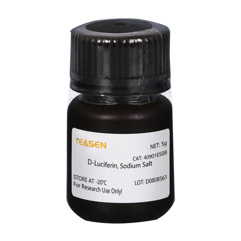
D-luciferine is een veelgebruikt substraat voor Luciferase en wordt veel gebruikt in de biotechnologie, met name in in vivo-beeldvormingstechnologie. Het werkingsmechanisme is dat luciferine (het substraat) wordt geoxideerd om licht uit te zenden als reactie op ATP en luciferase (zie onderstaande afbeelding). Het wordt geproduceerd wanneer er teveel luciferine aanwezig is. Het lichtkwantumgetal bleek positief gecorreleerd met de concentratie luciferase. Plasmiden die het gen dragen dat codeert voor luciferase (Luc), werden getransfecteerd in cellen en geïntroduceerd in proefdieren zoals ratten en muizen. Vervolgens werd in vivo fluoresceïne geïnjecteerd en werden veranderingen in de lichtintensiteit gedetecteerd met behulp van bioluminescentiebeeldvorming (BLI) om de voortgang van de ziekte of de werkzaamheid van geneesmiddelen in real time te monitoren, enzovoort. De invloed van ATP op het reactiesysteem kan ook worden gebruikt om energie of vitale functies aan te geven, afhankelijk van veranderingen in de intensiteit van de bioluminescentie.

D-luciferine wordt ook vaak gebruikt in in vitro-onderzoeken, waaronder luciferase- en ATP-niveauanalyse; reportergenanalyse; high-throughput sequencing en verschillende contaminatietesten. Er zijn momenteel drie productvormen: D-luciferine (vrij zuur), D-luciferinezouten (natrium- en kaliumzouten). Het belangrijkste verschil ligt in de oplosbaarheid: de eerste is oplosbaar in water en de oplosbaarheid van het buffersysteem is zwak, behalve oplosbaar in zwakke basen zoals NaOH- en KOH-oplossingen met een lage concentratie, oplosbaar in methanol en DMSO; de laatste is gemakkelijk oplosbaar in water of buffer, gemakkelijk te gebruiken, oplosmiddel niet-toxisch, vooral geschikt voor in vivo-experimenten. Als oplossing is er in de overgrote meerderheid van de toepassingen geen substantieel verschil tussen de drie producten.
Functies
- Geen straling, vrijwel ongevaarlijk voor levende organismen.
- Bioluminescentie, geen exciterende lichtbron.
- Zo gevoelig dat je het in een paar honderd cellen kunt detecteren.
- Goede penetratie, weefseldiepte van 3-4 cm is nog steeds te detecteren.
- Hoge signaal-ruisverhouding, sterk fluorescentiesignaal en goede anti-interferentie.
Sollicitatie
- Bij het tumorigenese-experiment bij naakte muizen werd de tumorgroei in realtime waargenomen zonder invasie, zonder dat de tumor werd afgestoten.
- Om het effect van de toediening op de tumorgroei of metastasering te testen, kan het fluoresceïnesubstraat binnen 3 uur volledig worden verwijderd, zonder dat het medicijnexperiment wordt verstoord.
- De lokalisatie en distributie van vreemde cellen bij dieren werden gedetecteerd.
- Het doelgen of de promotor van het doelgen wordt gefuseerd met het luciferasegen om veranderingen in de genexpressie te detecteren tijdens een medicijnbehandeling of ziekteprogressie.
- Toezicht op stamceltransplantatie, overleving en proliferatie; Volg de distributie en migratie van stamcellen in vivo.
Specificaties
| Engels synoniem | (S)-4,5-Dihydro-2-(6-hydroxy-2-benzothiazolyl)-4-thiazoolcarbonzuur zuur natrium zout; D-Luciferine vuurvlieg, natriumzout monohydraat; |
| CAS-NR. | 103404-75-7 |
| Formule | NaC11H7N2O3S2·H2O |
| Moleculair gewicht | 320,32 gram/mol |
| Verschijning | Lichtgeel poeder |
| Oplosbaarheid | Oplosbaar in water (100 mg/ml) |
| Zuiverheid (HPLC-test) | ≥95% |
Componenten
| Componenten nr. | Naam | 40901ES01 | 40901ES02 | 40901ES03 | 40901ES08 | 40901ES10 |
| 40901 | D-Luciferine, natriumzout | 100mg | 500mg | 1 gram | 5 gram | 10 gram |
Opslag
Het product moet worden bewaard bij -15℃ ~ -25℃ voor 1 jaar.
[1] Guo Y, Guo Y, Chen C, et al. Circ3823 draagt bij aan de groei, metastasering en angiogenese van colorectale kanker: betrokkenheid van de miR-30c-5p/TCF7-as. Mol Cancer. 2021;20(1):93. Gepubliceerd op 25 juni 2021. doi:10.1186/s12943-021-01372-0(IF:27.401)
[2] Wang Z, Yu L, Wang Y, et al. Dynamische aanpassing van niet-stralende en stralende verzwakking van AIE-moleculen versterkt NIR-II-beeldvorming gemedieerde fotothermische therapie en immunotherapie. Adv Sci (Weinh). 2022;9(8):e2104793. doi:10.1002/advs.202104793(IF:16.806)
[3] Liu Q, Sheng Z, Cheng C, et al. Anestheticum Propofol bevordert tumormetastase in longen via GABA<sub>A</sub> R-afhankelijke TRIM21-modulatie van Src-expressie. Adv Sci (Weinh). 2021;8(18):e2102079. doi:10.1002/advs.202102079(IF:16.806)
[4] Zhou M, Zhang J, Shen J, et al. Waterstofsulfide-gekoppelde persulfidatie van ABI4 controleert ABA-reacties via de transactivatie van MAPKKK18 in Arabidopsis. Mol Plant. 2021;14(6):921-936. doi:10.1016/j.molp.2021.03.007(IF:13.164)
[5] Wang J, Wang C, Li Y, et al. Potentieel van peptide-gemanipuleerde exosomen met overgeëxprimeerde miR-92b-3p in anti-angiogene therapie van eierstokkanker. Clin Transl Med. 2021;11(5):e425. doi:10.1002/ctm2.425(IF:11.492)
[6] Mao Z, Wei X, Li L, et al. Arabidopsis cryptochroom 1 controleert fotomorfogenese door regulatie van H2A.Z-afzetting. Plant Cell. 2021;33(6):1961-1979. doi:10.1093/plcell/koab091(IF:11.277)
[7] Xu P, Chen H, Li T, et al. Blauw licht-afhankelijke interacties van CRY1 met GID1 en DELLA-eiwitten reguleren gibberellinesignalering en fotomorfogenese in Arabidopsis. Plant Cell. 2021;33(7):2375-2394. doi:10.1093/plcell/koab124(IF:11.277)
[8] Lin S, Wen Z, Li S, et al. LncRNA Neat1 bevordert de ontstekingsreactie van macrofagen en fungeert als een therapeutisch doelwit bij door titaniumdeeltjes geïnduceerde osteolyse. Acta Biomater. 2022;142:345-360. doi:10.1016/j.actbio.2022.02.007(IF:8.947)
[9] Zhang Z, Zeng D, Zhang W, et al. Modulatie van oxidatieve fosforylering vergroot de antineoplastische activiteit van mitotische aurora-kinase-remming. Cell Death Dis. 2021;12(10):893. Gepubliceerd op 30 sep 2021. doi:10.1038/s41419-021-04190-w(IF:8.469)
[10] Wang L, Tong X, Zhou Z, et al. Circulair RNA hsa_circ_0008305 (circPTK2) remt TGF-β-geïnduceerde epitheliale-mesenchymale transitie en metastase door TIF1γ te controleren bij niet-kleincellige longkanker. Mol Cancer. 2018;17(1):140. Gepubliceerd op 27 sep 2018. doi:10.1186/s12943-018-0889-7(IF:7.776)
[11] Cao X, Xu P, Liu Y, et al.Cryptochroom 1 van Arabidopsis bevordert de stomatale ontwikkeling door onderdrukking van AGB1-remming van SPEECHLESS DNA-bindende activiteit. J Integr Plant Biol. 2021;63(11):1967-1981. doi:10.1111/jipb.13168(IF:7.061)
[12] Du SS, Li L, Li L, et al. Foto-geëxciteerde Cryptochrome2 interageert direct met TOE1 en TOE2 bij de regulatie van de bloei. Plant Physiol. 2020;184(1):487-505. doi:10.1104/pp.20.00486(IF:6.902)
[13] Zhou H, Qi Z, Pei P, et al. Biocompatibele nanomellen voor gevoelige detectie en fotodynamische therapie van kanker in een vroeg stadium. Biomater Sci. 2021;9(18):6227-6235. Gepubliceerd 14 sep 2021. doi:10.1039/d1bm00847a(IF:6.843)
[14] Zhang L, Zhou J, Yan Y, et al. Hulpstofvrije nanodispersie van 7-ethyl-10-hydroxycamptothecine oefent krachtige therapeutische effecten uit tegen pancreaskankercellijnen en van patiënten afkomstige xenograften. Cancer Lett. 2019;465:36-44. doi:10.1016/j.canlet.2019.08.019(IF:6.508)
[15] Geng Y, Duan H, Xu L, et al. BMP-2 en VEGF-A modRNAs in collageensteigers sturen synergetisch botherstel aan via osteogene en angiogene paden. Commun Biol. 2021;4(1):82. Gepubliceerd 19 jan. 2021. doi:10.1038/s42003-020-01606-9(IF:6.268)
[16] Qiu J, Peng P, Xin M, et al. ZBTB20-gemedieerde titaniumdeeltjes-geïnduceerde peri-implantaat osteolyse door het bevorderen van macrofaag ontstekingsreacties. Biomater Sci. 2020;8(11):3147-3163. doi:10.1039/d0bm00147c(IF:6.183)
[17] Zhou C, Wang G, Jing W, Tan X, Guo H. Antikankereigenschappen en mechanismen van enkelvoudig geprotoneerd dehydronorcantharidine-zilvercoördinatiepolymeer in een blaaskankermodel. Front Pharmacol. 2021;12:618668. Gepubliceerd op 23 februari 2021. doi:10.3389/fphar.2021.618668(IF:5.811)
[18] Sheng Y, Yu H, Pan H, et al. Genome-Wide Analysis van de genstructuur, expressie en eiwitinteracties van de perzik (Prunus persica) TIFY-genfamilie. Front Plant Sci. 2022;13:792802. Gepubliceerd 17 feb. 2022. doi:10.3389/fpls.2022.792802(IF:5.754)
[19] Jia MZ, Liu LY, Geng C, Jiang J. Activering van 1-aminocyclopropaan-1-carbonzuursynthasen bepaalt de stomatale dichtheid en de geclusterde verhouding op de bladepidermis van Arabidopsis als reactie op droogte. Front Plant Sci. 2021;12:758785. Gepubliceerd op 6 december 2021. doi:10.3389/fpls.2021.758785(IF:5.754)
[20] Xuan Z, Zhao L, Li Z, et al. EPS8L3 bevordert de proliferatie en metastasering van hepatocellulair carcinoom door de dimerisatie en internalisatie van EGFR te moduleren. Am J Cancer Res. 2020;10(1):60-77. Gepubliceerd op 1 januari 2020. (IF:5.177)
[21] Chen G, Fan XY, Zheng XP, Jin YL, Liu Y, Liu SC. Menselijke navelstreng-afgeleide mesenchymale stamcellen verbeteren insulineresistentie via PTEN-gemedieerde kruisspraak tussen de PI3K/Akt en Erk/MAPKs signaalpaden in de skeletspieren van db/db muizen. Stem Cell Res Ther. 2020;11(1):401. Gepubliceerd 2020 Sep 16. doi:10.1186/s13287-020-01865-7(IF:5.116)
[22] Chen G, Fan XY, Zheng XP, Jin YL, Liu Y, Liu SC. Menselijke navelstreng-afgeleide mesenchymale stamcellen verbeteren insulineresistentie via PTEN-gemedieerde kruisspraak tussen de PI3K/Akt en Erk/MAPKs signaalpaden in de skeletspieren van db/db muizen. Stem Cell Res Ther. 2020;11(1):401. Gepubliceerd 2020 Sep 16. doi:10.1186/s13287-020-01865-7(IF:5.116)
[23] You D, Du D, Zhao X, Li X, Ying M, Hu X. Mitochondriaal malaatenzym 2 bevordert borstkankermetastase via stabilisering van HIF-1α onder hypoxie. Chin J Cancer Res. 2021;33(3):308-322. doi:10.21147/j.issn.1000-9604.2021.03.03(IF:5.087)
[24] Qin L, Zhao R, Chen D, et al. Chimere antigeenreceptor-T-cellen die op PD-L1 zijn gericht, onderdrukken tumorgroei. Biomark Res. 2020;8:19. Gepubliceerd op 3 juni 2020. doi:10.1186/s40364-020-00198-0(IF:4.866)
[25] Zuo X, Xiang W, Li K, et al. MdGRF11, een groeiregulerende factor, neemt deel aan de regulatie van de bloeitijd en interageert met MdTFL1/MdFT1 in appel. Plant Sci. 2022;321:111339. doi:10.1016/j.plantsci.2022.111339(IF:4.729)
[26] Yang X, Yu Q, Xu H, Zhou J. Upregulatie van CD22 door Chidamide bevordert de functionaliteit van CAR T-cellen. Sci Rep. 2021;11(1):20637. Gepubliceerd 19 okt. 2021. doi:10.1038/s41598-021-00227-4(IF:4.380)
[27] Mei Z, Zhang K, Lam AK, et al. MUC1 als doelwit voor CAR-T-therapie bij plaveiselcelcarcinoom van hoofd en hals. Cancer Med. 2020;9(2):640-652. doi:10.1002/cam4.2733(IF:3.357)
[28] Zhang Z, Liu L, Cao S, Zhu Y, Mei Q. Genlevering van TIPE2 remt de ontwikkeling en metastasering van borstkanker via CD8+ T- en NK-cel-gemedieerde antitumorreacties. Mol Immunol. 2017;85:230-237. doi:10.1016/j.molimm.2017.03.007(IF:3.236)
[29] Yuan L, Zhou M, Huang D, et al. Resveratrol remt de invasie en metastasering van colonkanker door omkering van de epitheliale-mesenchymale transitie via de AKT/GSK‑3β/Snail-signaalroute. Mol Med Rep. 2019;20(3):2783-2795. doi:10.3892/mmr.2019.10528(IF:2.952)
[30] Zhang C, Liu X, Chen F, et al. Gliotoxine vernietigt de barrièrefunctie van het longepitheel door de vorming van cofiline-oligomeren te verminderen om de oplosbaarheid van actinestressvezels te bevorderen. Microb Pathog. 2018;123:169-176. doi:10.1016/j.micpath.2018.07.007(IF:2.332)
Storage
FAQ
- Thiazoline-4-carboxylic acid.
1) Solubility: The salt form is easily soluble in water. The solubility of potassium salt is 60mg/ml, and that of sodium salt is 100mg/ml. Free acid is not easily soluble in water. It can be dissolved by a weak alkaline solution of sodium bicarbonate, and its solubility in methanol is 10mg/ml, and in DMSO is 50mg/ml.
2) Toxicity: The salt form is more convenient to use during the process, especially in in vivo imaging experiments, as it can dissolve in water and thus has less reaction toxicity (Fluorescein is a low-molecular-weight organic compound composed of phenylthiazole and thiazole carboxylic acid groups, which has low toxicity). 3) Usage effect: There is no significant difference. In in vivo experimental studies, the usage rate of potassium salt is higher.
1. Instruments with a biochemiluminescence detection module. The light produced by luciferin can be detected by a photometer or a scintillation counter. Common in vivo imaging instruments: such as the IVIS® Lumina small animal in vivo imaging system, and the In-Vivo Xtreme multi-mode small animal in vivo imaging instrument from Bruker, Germany.
2. Multi-functional microplate readers: It is necessary to confirm with the instrument manufacturer whether it is a specific biochemiluminescence detection module (note: cannot use a fluorescence microscope).
1) Injection method: It can be done through intraperitoneal injection or tail vein injection.
2) Injection volume: The scientific approach is to evaluate the injection dose based on the kinetic curve. The initial attempted injection dose was set at 150mg substrate/kg of mouse body weight. Therefore, the purchase quantity can be calculated according to the above method: If there are 10 mice, 22 - 25g, then 33 - 37.5mg of substrate is required.
Citations
Documents
| Manual |
|---|
| 40901ES_Manual_Ver.EN20250520.pdf |
| MSDS |
|---|
| 40901-MSDS-HB251208.pdf |
| Brochure |
|---|
| In_vivo_imaging_Leaving_Tumor_Cells_Nowhere_to_Hide.pdf |


Misschien vind je het ook leuk
Navraag
FAQ
Het product is alleen bedoeld voor onderzoeksdoeleinden en is niet bedoeld voor therapeutisch of diagnostisch gebruik bij mensen of dieren. Producten en inhoud worden beschermd door patenten, handelsmerken en auteursrechten die eigendom zijn van
Voor bepaalde toepassingen zijn mogelijk aanvullende intellectuele eigendomsrechten van derden vereist.