


Frequently Bought Together
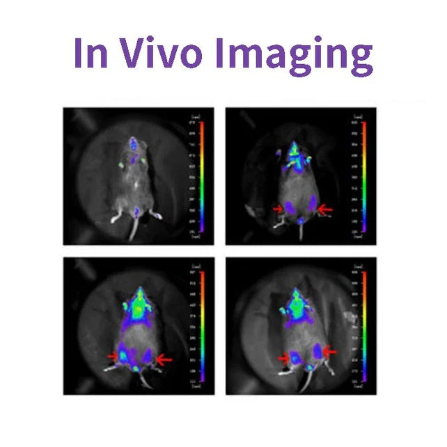
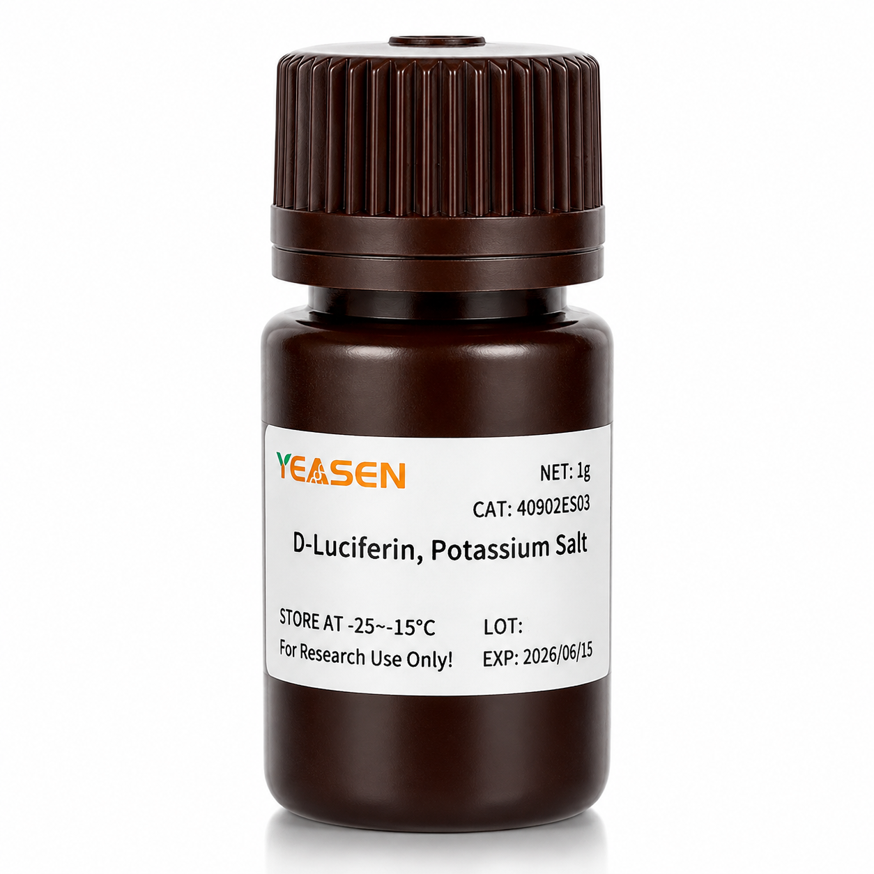
D-Luciferin ist ein häufiges Substrat für Luciferase und wird in der gesamten Biotechnologie, insbesondere in der In-vivo-Bildgebungstechnologie, häufig verwendet. Der Wirkungsmechanismus besteht darin, dass Luciferin (Substrat) in Gegenwart von ATP und Luciferase oxidiert werden kann, um Licht zu emittieren. Bei einem Überschuss an Fluorescein ist die Quantenzahl des erzeugten Lichts gleich der Luciferase-Konzentration, die positiv korreliert war (siehe Abbildung unten). Plasmide, die das für Luciferase kodierende Gen (Luc) tragen, wurden in Zellen transfiziert und in Versuchstiere wie Ratten und Mäuse eingeführt.

D-Luciferin wird auch häufig in In-vitro-Studien verwendet, einschließlich der Analyse von Luciferase- und ATP-Spiegeln, Reportergenanalyse, Hochdurchsatzsequenzierung und verschiedenen Kontaminationstests. Es gibt drei Produktformen: D-Luciferin (freie Säure), D-Luciferinsalz (Natriumsalz und Kaliumsalz). Der Hauptunterschied liegt in der Löslichkeit: Wasserlöslichkeit der ersteren und Löslichkeit des Puffersystems. Sie sind schwach löslich, außer in schwachen Basen wie niedrigen Konzentrationen von NaOH- und KOH-Lösungen. Löslich in Methanol und DMSO; Letzteres kann leicht in Wasser oder Puffer gelöst werden und ist einfach zu verwenden.
Merkmale
- Keine Strahlung, nahezu unschädlich für Lebewesen.
- Biolumineszenz, keine Anregungslichtquelle.
- So empfindlich, dass Sie es in einigen hundert Zellen erkennen können.
- Gute Penetration, 3-4cm Gewebetiefe noch erkennbar.
- Hohes Signal-Rausch-Verhältnis, starkes Fluoreszenzsignal und gute Entstörungsfähigkeit.
Anwendungen
- Im Tumorigenese-Experiment an Nacktmäusen wurde das Tumorwachstum ohne Invasion in Echtzeit, ohne Tumor-Stripping-Messung, beobachtet.
- Um die Wirkung der Verabreichung auf das Tumorwachstum oder die Metastasierung zu testen, kann das Fluoresceinsubstrat innerhalb von 3 Stunden vollständig eliminiert werden, ohne den Arzneimittelversuch zu beeinträchtigen.
- Die Lokalisierung und Verteilung fremder Zellen im Tier wurde nachgewiesen.
- Das Zielgen oder der Promotor des Zielgens wird mit dem Luciferase-Gen fusioniert, um Änderungen der Genexpression während der Arzneimittelbehandlung oder des Krankheitsverlaufs zu erkennen.
- Überwachung der Stammzelltransplantation, des Überlebens und der Proliferation; Verfolgung der Verteilung und Migration von Stammzellen in vivo.
Technische Daten
| Englisches Synonym | (S)-4,5-Dihydro-2-(6-hydroxy-2-benzothiazolyl)-4-thiazolcarbonsäure Kaliumsalz; D-Luciferin-Glühwürmchen, Kaliumsalz |
| CAS-NR. | 115144-35-9 |
| Formel | C11H7N2O3S2K |
| Molekulargewicht | 318,42 g/mol |
| Aussehen | Hellgelbes Pulver |
| Löslichkeit | Wasserlöslich(60 mg/ml) |
Komponenten
| Komponenten Nr. | Name | 40902ES01 | 40902ES02 | 40902ES03 | 40902ES08 |
| 40902 | D-Luciferin, Kaliumsalz | 100 mg | 500 mg | 1 g | 5 g |
Zahlen

Zitiert aus „Ein neuartiger allosterischer Inhibitor der Phosphoglycerat-Mutase 1 unterdrückt Wachstum und Metastasierung von nicht-kleinzelligem Lungenkrebs“, Cell Metab.2019;30(6):1107-1119
Versand und Lagerung
Das Produkt wird mit Trockeneis geliefert und kann bei -15℃ gelagert werden ~ -25℃ für 1 Jahr.
[1] Gu Y, Wang Y, He L, et al. Zirkuläre RNA circIPO11 treibt die Selbsterneuerung von Leberkrebs-initiierenden Zellen über Hedgehog-Signale voran. Mol Cancer. 2021;20(1):132. Veröffentlicht am 14. Oktober 2021. doi:10.1186/s12943-021-01435-2(IF:27.401)
[2] Huang K, Liang Q, Zhou Y, et al. Ein neuartiger allosterischer Inhibitor der Phosphoglycerat-Mutase 1 unterdrückt Wachstum und Metastasierung von nicht-kleinzelligem Lungenkrebs [veröffentlichte Korrektur erscheint in Cell Metab. 2021 Jan 5;33(1):223]. Cell Metab. 2019;30(6):1107-1119.e8. doi:10.1016/j.cmet.2019.09.014(IF:22.415)
[3] Dong X, Cheng R, Zhu S, et al. Ein WO<sub>2.9</sub>-WSe<sub>2</sub>-Nanoradiosensitizer mit Heteroübergangsstruktur verbessert die lokale Tumorablation und die Checkpoint-Blockade-Immuntherapie bei niedriger Strahlungsdosis. ACS Nano. 2020;14(5):5400-5416. doi:10.1021/acsnano.9b08962(IF:14.588)
[4] Zheng DW, Gao F, Cheng Q, et al. Ein Impfstoff-basiertes Nanosystem zur Initiierung der angeborenen Immunität und Verbesserung der Tumorimmuntherapie. Nat Commun. 2020;11(1):1985. Veröffentlicht 2020 Apr 24. doi:10.1038/s41467-020-15927-0(IF:12.121)
[5] Hu J, Su H, Cao H, et al. AUXIN RESPONSE FACTOR7 integriert Gibberellin- und Auxin-Signalisierung über Interaktionen zwischen DELLA- und AUX/IAA-Proteinen, um die Kambialaktivität in Pappeln zu regulieren. Plant Cell. 2022;34(7):2688-2707. doi:10.1093/plcell/koac107(IF:11.277)
[6] Qi P, Huang M, Hu X, et al. Ein Ralstonia solanacearum-Effektor zielt auf TGA-Transkriptionsfaktoren ab, um die Salicylsäure-Signalübertragung zu untergraben. Pflanzenzelle. 2022;34(5):1666-1683. doi:10.1093/plcell/koac015(IF:11.277)
[7] Huang X, Qiu M, Wang T, et al. Trägerfreie multifunktionale Nanomedizin zur intraperitonealen disseminierten Eierstockkrebstherapie. J Nanobiotechnology. 2022;20(1):93. Veröffentlicht am 22. Februar 2022. doi:10.1186/s12951-022-01300-4(IF:10.435)
[8] Tang Y, Lin S, Yin S, et al. In-situ-Gasschäumen basierend auf Magnesiumpartikelabbau: Ein neuartiger Ansatz zur Herstellung injizierbarer makroporöser Hydrogele. Biomaterialien. 2020;232:119727. doi:10.1016/j.biomaterials.2019.119727(IF:10.273)
[9] Zhang X, Zheng S, Hu C, et al. Krebsassoziierte Fibroblasten-induzierte lncRNA UPK1A-AS1 verleiht Platinresistenz bei Bauchspeicheldrüsenkrebs durch effiziente Reparatur von Doppelstrangbrüchen. Oncogene. 2022;41(16):2372-2389. doi:10.1038/s41388-022-02253-6(IF:9.867)
[10] Qiao K, Liu Y, Xu Z, et al. RNA m6A-Methylierung fördert die Bildung von vaskulogener Mimikry bei hepatozellulärem Karzinom über den Hippo-Signalweg. Angiogenese. 2021;24(1):83-96. doi:10.1007/s10456-020-09744-8(IF:9.780)
[11] Wu Q, Kuang K, Lyu M, et al. Allosterische Deaktivierung von PIFs und EIN3 durch Mikroproteine bei der Lichtkontrolle der Pflanzenentwicklung. Proc Natl Acad Sci US A. 2020;117(31):18858-18868. doi:10.1073/pnas.2002313117(IF:9.412)
[12] Meng DF, Sun R, Liu GY, et al. S100A14 unterdrückt die Metastasierung von Nasopharynxkarzinomen durch Hemmung der NF-kB-Signalgebung durch Abbau von IRAK1. Oncogene. 2020;39(30):5307-5322.doi:10.1038/s41388-020-1363-8(IF:7.971)
[13] Gao Y, Wang J, Han H, et al. Ein nanopartikelhaltiges Polycaprolacton-Implantat zur Bekämpfung des Wiederauftretens von Brustkrebs nach Resektion. Nanoskala. 2021;13(34):14417-14425. Veröffentlicht am 2. September 2021. doi:10.1039/d1nr04125h(IF:7.790)
[14] Jiang W, Li T, Guo J, et al. Bispezifische c-Met/PD-L1 CAR-T-Zellen haben eine verstärkte therapeutische Wirkung auf hepatozelluläres Karzinom. Front Oncol. 2021;11:546586. Veröffentlicht am 10. März 2021. doi:10.3389/fonc.2021.546586(IF:6.244)
[15] Zhou Y , Zhou C , Zou Y , et al. Multi-pH-sensitive Polymer-Wirkstoff-Konjugat-Mischmizellen für die effiziente gemeinsame Verabreichung von Doxorubicin und Curcumin zur synergistischen Unterdrückung der Tumormetastasierung. Biomater Sci. 2020;8(18):5029-5046. doi:10.1039/d0bm00840k(IF:6.183)
[16] Zhao X, Liu X, Zhang P, et al. Injizierbares Peptid-Hydrogel als intraperitoneales Triptolid-Depot zur Behandlung von orthotopem hepatozellulärem Karzinom. Acta Pharm Sin B. 2019;9(5):1050-1060. doi:10.1016/j.apsb.2019.06.001(IF:5.808)
[17] Li M, Wang J, Yu Y, et al. Charakterisierung von mesenchymalen Stammzellen, die aus Bisphosphonat-bedingter Osteonekrose des Zahnfleisches von Kieferpatienten gewonnen wurden. Stem Cell Rev Rep. 2022;18(1):378-394. doi:10.1007/s12015-021-10241-8(IF:5.739)
[18] Zhang X, Li Y, Ji J, et al. Gadd45g initiiert die Differenzierung embryonaler Stammzellen und hemmt die Brustkrebsentstehung. Cell Death Discov. 2021;7(1):271. Veröffentlicht am 2. Oktober 2021. doi:10.1038/s41420-021-00667-x(IF:5.241)
[19] Wu D, Lv J, Zhao R, et al. PSCA ist ein Ziel chimärer Antigenrezeptor-T-Zellen bei Magenkrebs. Biomark Res. 2020;8:3. Veröffentlicht am 28. Januar 2020. doi:10.1186/s40364-020-0183-x(IF:4.866)
[20] Qiao K, Chen C, Liu H, Qin Y, Liu H. Pinin induziert den epithelialen-mesenchymalen Übergang beim hepatozellulären Karzinom durch Regulierung der m6A-Modifikation. J Oncol. 2021;2021:7529164. Veröffentlicht am 7. Dezember 2021. doi:10.1155/2021/7529164(IF:4.375)
[21] Li K, Liu T, Chen J, Ni H, Li W. Survivin in aus Brustkrebs stammenden Exosomen aktiviert Fibroblasten durch Hochregulierung von SOD1, dessen Rückkopplung die Krebsproliferation und Metastasierung fördert. J Biol Chem. 2020;295(40):13737-13752. doi:10.1074/jbc.RA120.013805(IF:4.238)
[22] Kong Y, Feng Z, Chen A, et al. Das natürliche Flavonoid Galangin löst Apoptose, Pyroptose und Autophagie im Glioblastom aus. Front Oncol. 2019;9:942. Veröffentlicht am 27. September 2019. doi:10.3389/fonc.2019.00942(IF:4.137)
[23] Kong Y, Feng Z, Chen A, et al. Das natürliche Flavonoid Galangin löst Apoptose, Pyroptose und Autophagie im Glioblastom aus. Front Oncol. 2019;9:942. Veröffentlicht am 27. September 2019. doi:10.3389/fonc.2019.00942(IF:4.137)
[24] Hu J, Wang Y, Yuan Y. Inhibitoren der APE1-Redoxfunktion hemmen wirksam die Replikation des γ-Herpesvirus in vitro und in vivo. Antiviral Res. 2021;185:104985. doi:10.1016/j.antiviral.2020.104985(IF:4.101)
[25] Li Q, Wei D, Feng F, et al. α2,6-gebundene Sialinsäure dient als hochaffiner Rezeptor für die onkolytische Virotherapie mit dem Newcastle-Disease-Virus. J Cancer Res Clin Oncol. 2017;143(11):2171-2181. doi:10.1007/s00432-017-2470-y(IF:3.503)
[26] Han B, Jiang P, Liu W, et al. Rolle von Daucosterol Linoleat bei Brustkrebs: Studien zu Apoptose und Metastasierung. J Agric Food Chem. 2018;66(24):6031-6041. doi:10.1021/acs.jafc.8b01387(IF:3.412)
[27] Hu H, Zhang Z, Wang R, et al. Die BGC823-Zelllinie mit der stabilen Expression von iRFP720 behält ihre primären Eigenschaften mit vielversprechender Fluoreszenzbildgebungsfähigkeit. DNA Cell Biol. 2020;39(5):900-908. doi:10.1089/dna.2019.5057(IF:3.314)
[28] Ni XR, Zhao YY, Cai HP, et al.Auf Transferrinrezeptor 1 ausgerichtete optische Bildgebung zur Identifizierung des Gliomrands in Mausmodellen. J Neurooncol. 2020;148(2):245-258. doi:10.1007/s11060-020-03527-3(IF:3.267)
[29] Feng H, Tang J, Zhang P, Miao Y, Wu T, Cheng Z. Antiadipogene 18,19-Seco-Ursan-Stereoisomere und Olean-artige Saponine aus Ilex cornuta-Blättern. Phytochemie. 2020;175:112363. doi:10.1016/j.phytochem.2020.112363(IF:3.044)
[30] Zhao Y, Li T, Tian S, et al. Effektive Hemmung von MYC-amplifiziertem Medulloblastom der Gruppe 3 durch gezieltes EIF4A1. Cancer Manag Res. 2020;12:12473-12485. Veröffentlicht am 3. Dezember 2020. doi:10.2147/CMAR.S278844(IF:2.886)
Storage
FAQ
1) Solubility: The salt form is easily soluble in water. The solubility of potassium salt is 60mg/ml, and that of sodium salt is 100mg/ml. Free acid is not easily soluble in water. It can be dissolved by a weak alkaline solution of sodium bicarbonate, and its solubility in methanol is 10mg/ml, and in DMSO is 50mg/ml.
2) Toxicity: The salt form is more convenient to use during the process, especially in in vivo imaging experiments, as it can dissolve in water and the reaction toxicity will be smaller (Fluorescein is a low-molecular-weight organic compound composed of phenylthiazole and thiazole carboxyl group, with low toxicity).
3) Usage effect: There is no significant difference. In in vivo experimental studies, the usage rate of potassium salt is higher.
2. Native coelenterazine is an action substrate for various luciferases such as sea kidney luciferase (Rluc) and Gaussia luciferase (Gluc).
1. Instruments with a biochemiluminescence detection module. The light produced by luciferin can be detected by a photometer or a scintillation counter. Common in vivo imaging instruments: such as the IVIS® Lumina small animal in vivo imaging system, the In-Vivo Xtreme multi-mode small animal in vivo imaging instrument from Bruker, Germany.
2. Multi-functional microplate readers: It is necessary to confirm with the instrument manufacturer whether it is a specific biochemiluminescence detection module (note: cannot use a fluorescence microscope).
2. The cells themselves also contain a certain amount of ATP and magnesium, which can promote the reaction. In most cases, the ATP and magnesium contained in the cells are sufficient for the reaction of D-fluorescein salt, but the content varies depending on the type of cells. You can first try using a culture medium without additional ATP and magnesium, and conduct the experiment normally to see the results.
1) Injection method: It can be done through intraperitoneal injection or tail vein injection.
2) Injection volume: The scientific approach is to evaluate the injection dose based on the kinetic curve. The initial attempted injection dose is: 150mg substrate/kg of mouse body weight. Therefore, the purchase quantity can be calculated according to the above method: If there are 10 mice, 22 - 25g, then 33 - 37.5mg of substrate is required.
Citations
Documents
| Brochure |
|---|
| In_vivo_imaging_Leaving_Tumor_Cells_Nowhere_to_Hide.pdf |
Applications


Sie können auch mögen
Anfrage
FAQ
Das Produkt ist nur für Forschungszwecke bestimmt und nicht für die therapeutische oder diagnostische Anwendung bei Menschen oder Tieren. Produkte und Inhalte sind durch Patente, Marken und Urheberrechte von
Für bestimmte Anwendungen sind möglicherweise zusätzliche geistige Eigentumsrechte Dritter erforderlich.