


Description
The MycAway™ Plus-Color One-Step Mycoplasma Detection Kit is a rapid detection product for mycoplasma contamination in cell culture which was developed based on the unique isothermal amplification technology of YEASEN. Compared with the previous mycoplasma detection products, this kit is optimized and has greatly reduced the false positive rate, improved the accuracy of the test, and enhanced the identification of negative and positive results. At the end of amplification, the sample can be left at room temperature for a period of time without the change from negative to positive, and thus will not affect the judgment of the results.
The principle is that if the cell culture is contaminated with mycoplasma, the conserved sequences of mycoplasma DNA will be amplified heavily and rapidly, causing the color of the reaction solution to change from blue-purple to sky-blue, and the result is visible to the naked eye, without electrophoresis.
MycAway™ Plus-Color One-Step Mycoplasma Detection Kit can detect multiple mycoplasma species, including eight that are common in cell culture. The traditional nested PCR method for mycoplasma detection is susceptible to influence by the inhibitors in cell culture supernatant and showed a false-negative result. Also if needed to perform the electrophoresis, cap opening may increase the contamination and lead to a false-positive result. The one-step mycoplasma detection kit has none of these disadvantages, also its detection sensitivity and accuracy are much higher than those of the PCR method.
Yeasen offers comprehensive solutions for Mycoplasma contamination. Explore our related products: Concept of Mycoplasma and the Impact of Contamination
Feature
- The one-step operation results in less than 60 minutes
- Use a water bath or PCR instrument to test
- 22 mycoplasma species were accurately detected by the kit, including 8 common species
- Results just identified by the visual inspection
Application
- Mycoplasma Detection
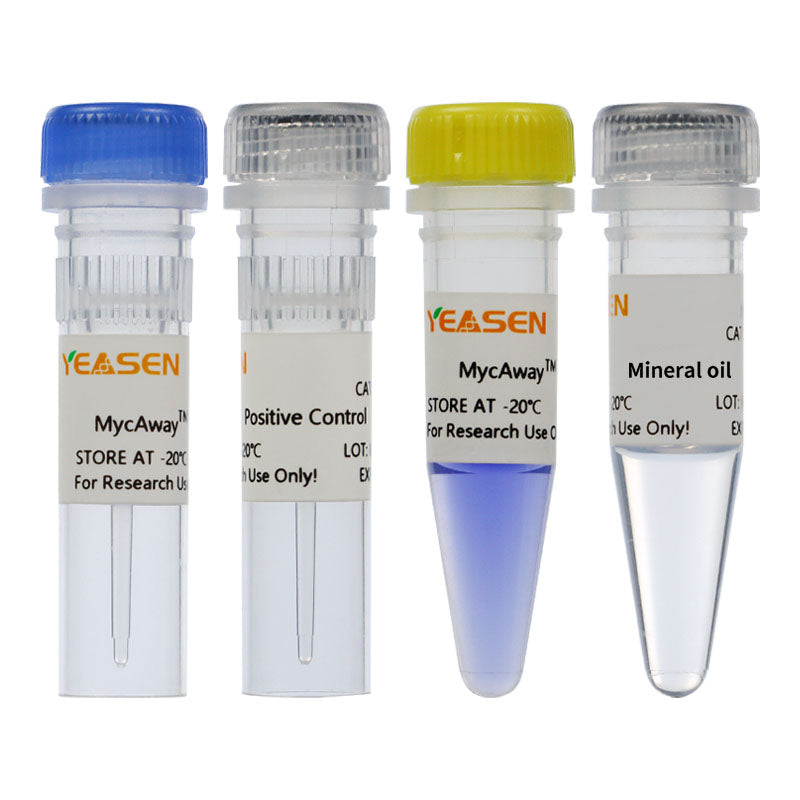
Components
| Components No. | Name | 40612ES25 (25 T) | 40612ES60 (100 T) |
| 40612-A | MycAway™ -Color Lamp | 425 μL | 4 × 425 μL |
| 40612-B | MycAway™-Color Lamp Primer | 50 μL | 4 × 50 μL |
| 40612-C | Positive Control | 10 μL | 4 × 10 μL |
| 40612-D | Mineral oil | 500 μL | 4 × 500 μL |
Shipping and Storage
All the components are shipped with dry ice and can be stored at -15℃ ~ -25℃ for 18 months. Please keep away from the light.
Figures
- The detection procedure
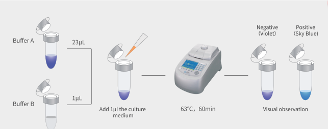
Figure 1. The detection procedure by MycAway™ Plus–Color One-Step Mycoplasma Detection Kit from YEASEN

FAQ
Q: What is the detection principle of the 40612 one-step rapid mycoplasma test kit (anti-contamination version)?
A: This kit contains UDG enzyme to remove the aerosol contamination of PCR product containing dU, which greatly inhibits false positive reactions and improves the accuracy of the test. Moreover, the added UDG enzyme functions at room temperature and is temperature-sensitive and prone to inactivation, without affecting the experimental results. If the cell culture is contaminated by mycoplasma, the conserved sequences of mycoplasma DNA will be amplified in large quantities and rapidly, causing the reaction solution to change from blue-purple to sky-blue. The result can be easily identified by the naked eye and no electrophoresis is required.
Q: How to use the one-step rapid mycoplasma detection kit (anti-contamination version)?
A: This reagent kit is much simpler to use. Just take 1 μL of the suspected mycoplasma-contaminated cell culture supernatant and add it to the reaction system. First, place the sample at 25-37 ℃ for 5 minutes, then incubate it at a constant temperature of 63 °C in a water bath or PCR instrument for 60 minutes. Observe that the reaction solution changes from blue-purple to sky-blue, and the result can be clearly seen without the need for electrophoresis.
Q: Using the 40612 one-step mycoplasma detection kit, we tested for the presence of mycoplasma contamination in organic solvents such as DMSO and ethanol. We found that the experimental group showed a sky-blue color. Does this mean that there is mycoplasma contamination in these organic solvents?
A: The mycoplasma kit is unstable and prone to degradation in ethanol, DMSO organic solvents, and in environments with strong acids and strong bases. As a result, the color changes to a sky blue. This kit can only be used for cell detection and should not be used in organic solvents or in environments with strong acids and strong bases for detection.
Q: I tested the liquid inside this. After adding it, the color in the PCR tube immediately turned sky blue. What's the reason for this? When testing pancreatic enzymes - EDTA, the color changed directly after adding the sample?
A: 40612 Principle: Mg2++HNB = Blue color. As PCR increases, pyrophosphate accumulates continuously. Pyrophosphate combines with Mg2+, while HNB remains free, resulting in a sky blue color. This reagent contains sodium dihydrogen phosphate, which may be the reason for the immediate chemical reaction between this ion and Mg2+ causing the reaction to occur and change color. EDTA can chelate calcium and magnesium ions, thereby accelerating the color change reaction.
Q: What is the concentration of the positive control?
A: 10 to the power of 5 copies per ul.
Q: Is it a mycoplasma in the positive control?
A: No, it's a linearized segment.
Documents
Citations & References:
[1] Shi N, Yang Q, Zhang H, et al. Restoration of dystrophin expression in mice by suppressing a nonsense mutation through the incorporation of unnatural amino acids. Nat Biomed Eng. 2022;6(2):195-206. doi:10.1038/s41551-021-00774-1(IF:25.671)
[2] Zhang S, Yu F, Che A, et al. Neuroendocrine Regulation of Stress-Induced T Cell Dysfunction during Lung Cancer Immunosurveillance via the Kisspeptin/GPR54 Signaling Pathway. Adv Sci (Weinh). 2022;9(13):e2104132. doi:10.1002/advs.202104132(IF:16.806)
[3] Li Y, Xue B, Zhang M, et al. Transcription-coupled structural dynamics of topologically associating domains regulate replication origin efficiency. Genome Biol. 2021;22(1):206. Published 2021 Jul 12. doi:10.1186/s13059-021-02424-w(IF:13.583)
[4] Wu L, Xu Y, Zhao H, et al. FcγRIIB potentiates differentiation of myeloid-derived suppressor cells to mediate tumor immunoescape. Theranostics. 2022;12(2):842-858. Published 2022 Jan 1. doi:10.7150/thno.66575(IF:11.556)
[5] Tan B, Shi X, Zhang J, et al. Inhibition of Rspo-Lgr4 Facilitates Checkpoint Blockade Therapy by Switching Macrophage Polarization. Cancer Res. 2018;78(17):4929-4942. doi:10.1158/0008-5472.CAN-18-0152(IF:9.130)
[6] Yan G, Zhao H, Zhang Q, et al. A RIPK3-PGE<sub>2</sub> Circuit Mediates Myeloid-Derived Suppressor Cell-Potentiated Colorectal Carcinogenesis. Cancer Res. 2018;78(19):5586-5599. doi:10.1158/0008-5472.CAN-17-3962(IF:9.130)
[7] Gu Z, Shi C, Li J, et al. Palbociclib-based high-throughput combination drug screening identifies synergistic therapeutic options in HPV-negative head and neck squamous cell carcinoma. BMC Med. 2022;20(1):175. Published 2022 May 12. doi:10.1186/s12916-022-02373-6(IF:8.775)
[8] Wu L, Zhang X, Zheng L, et al. RIPK3 Orchestrates Fatty Acid Metabolism in Tumor-Associated Macrophages and Hepatocarcinogenesis. Cancer Immunol Res. 2020;8(5):710-721. doi:10.1158/2326-6066.CIR-19-0261(IF:8.728)
[9] Qin J, Zhang X, Tan B, et al. Blocking P2X7-Mediated Macrophage Polarization Overcomes Treatment Resistance in Lung Cancer. Cancer Immunol Res. 2020;8(11):1426-1439. doi:10.1158/2326-6066.CIR-20-0123(IF:8.728)
[10] Cao M, Huang W, Chen Y, et al. Chronic restraint stress promotes the mobilization and recruitment of myeloid-derived suppressor cells through β-adrenergic-activated CXCL5-CXCR2-Erk signaling cascades. Int J Cancer. 2021;149(2):460-472. doi:10.1002/ijc.33552(IF:7.396)
[11] Yao R, Alkhawtani AYF, Chen R, Luan J, Xu M. Rapid and efficient in vivo angiogenesis directed by electro-assisted bioprinting of alginate/collagen microspheres with human umbilical vein endothelial cell coating layer [published correction appears in Int J Bioprint. 2020 Sep 17;6(4):309]. Int J Bioprint. 2019;5(2.1):194. Published 2019 Jun 24. doi:10.18063/ijb.v5i2.1.194(IF:6.638)
[12] Liu L, Deng Y, Zheng Z, et al. Hsp90 Inhibitor STA9090 Sensitizes Hepatocellular Carcinoma to Hyperthermia-Induced DNA Damage by Suppressing DNA-PKcs Protein Stability and mRNA Transcription. Mol Cancer Ther. 2021;20(10):1880-1892. doi:10.1158/1535-7163.MCT-21-0215(IF:6.261)
[13] Wu S, Yang X, Tang W, et al. Chemotherapeutic Risk lncRNA-PVT1 SNP Sensitizes Metastatic Colorectal Cancer to FOLFOX Regimen. Front Oncol. 2022;12:808889. Published 2022 Mar 31. doi:10.3389/fonc.2022.808889(IF:6.244)
[14] Wu Q, Xuan YF, Su AL, Bao XB, Miao ZH, Wang YQ. TNKS inhibitors potentiate proliferative inhibition of BET inhibitors via reducing β-Catenin in colorectal cancer cells. Am J Cancer Res. 2022;12(3):1069-1087. Published 2022 Mar 15. (IF:6.166)
[15] Wu L, Zhao KQ, Wang W, et al. Nuclear receptor coactivator 6 promotes HTR-8/SVneo cell invasion and migration by activating NF-κB-mediated MMP9 transcription. Cell Prolif. 2020;53(9):e12876. doi:10.1111/cpr.12876(IF:5.753)
[16] Wei Z, Wang Y, Peng J, et al. CircRFWD3 promotes HNSCC metastasis by modulating miR-27a/b/PPARγ signaling. Cell Death Discov. 2022;8(1):285. Published 2022 Jun 11. doi:10.1038/s41420-022-01066-6(IF:5.241)
[17] Chen W, Weng Z, Xie Z, et al. Sequencing of methylase-accessible regions in integral circular extrachromosomal DNA reveals differences in chromatin structure. Epigenetics Chromatin. 2021;14(1):40. Published 2021 Aug 23. doi:10.1186/s13072-021-00416-5(IF:4.954)
[18] Wu Z, Zheng M, Zhang Y, et al. Hsa_circ_0043278 functions as competitive endogenous RNA to enhance glioblastoma multiforme progression by sponging miR-638. Aging (Albany NY). 2020;12(21):21114-21128. doi:10.18632/aging.103603(IF:4.831)
[19] Wang L, Zhou Y, Cao C, et al. The exon 12-containing LHX6 isoforms promote cervical cancer cell proliferation by regulating the MAPK signaling pathway [published online ahead of print, 2022 Apr 5]. Cancer Med. 2022;10.1002/cam4.4734. doi:10.1002/cam4.4734(IF:4.452)
[20] Zhang Q, Yan G, Lei J, et al. The SP1-12LOX axis promotes chemoresistance and metastasis of ovarian cancer. Mol Med. 2020;26(1):39. Published 2020 May 6. doi:10.1186/s10020-020-00174-2(IF:4.096)
[21] Hu J, Wu Q, Wang Z, et al. Inhibition of CACNA1H attenuates doxorubicin-induced acute cardiotoxicity by affecting endoplasmic reticulum stress. Biomed Pharmacother. 2019;120:109475. doi:10.1016/j.biopha.2019.109475(IF:3.743)
[22] Wang T, Lin F, Sun X, et al. HOXB8 enhances the proliferation and metastasis of colorectal cancer cells by promoting EMT via STAT3 activation. Cancer Cell Int. 2019;19:3. Published 2019 Jan 3. doi:10.1186/s12935-018-0717-6(IF:3.439)
[23] Meng LL, Wang JL, Xu SP, et al. Low serum gastrin associated with ER<sup>+</sup> breast cancer development via inactivation of CCKBR/ERK/P65 signaling. BMC Cancer. 2018;18(1):824. Published 2018 Aug 16. doi:10.1186/s12885-018-4717-7(IF:3.288)
[24] Ma J, Liu X, Liu P, et al. Identification of a new p53 responsive element in the promoter region of anillin. Int J Mol Med. 2020;45(5):1563-1570. doi:10.3892/ijmm.2020.4527(IF:3.098)
[25] Chen L, Chen L, Wan L, et al. Matrine improves skeletal muscle atrophy by inhibiting E3 ubiquitin ligases and activating the Akt/mTOR/FoxO3α signaling pathway in C2C12 myotubes and mice. Oncol Rep. 2019;42(2):479-494. doi:10.3892/or.2019.7205(IF:3.041)
[26] Men XM, Xu ZW, Tao X, Deng B, Qi KK. FNDC5 expression closely correlates with muscle fiber types in porcine longissimus dorsi muscle and regulates myosin heavy chains (MyHCs) mRNA expression in C2C12 cells. PeerJ. 2021;9:e11065. Published 2021 Apr 19. doi:10.7717/peerj.11065(IF:2.984)
[27] Zhao YQ, Wu T, Wang LF, et al. Targeting MUC1-C reverses the cisplatin resistance of esophageal squamous cell carcinoma in vitro and in vivo. Transl Cancer Res. 2021;10(2):645-655. doi:10.21037/tcr-20-2495(IF:1.241)
Related blog:
Payment & Security
Your payment information is processed securely. We do not store credit card details nor have access to your credit card information.
You may also like
Inquiry
FAQ
The product is for research purposes only and is not intended for therapeutic or diagnostic use in humans or animals. Products and content are protected by patents, trademarks, and copyrights owned by Yeasen Biotechnology. Trademark symbols indicate the country of origin, not necessarily registration in all regions.
Certain applications may require additional third-party intellectual property rights.
Yeasen is dedicated to ethical science, believing our research should address critical questions while ensuring safety and ethical standards.