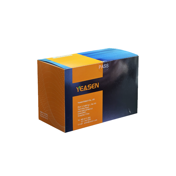
Description
The Annexin V-YSFluor™ 488/PI Apoptosis Detection Kit utilizes Annexin V conjugated to YSFluor™ 488 as a fluorescent probe to detect early-stage apoptosis. The assay can be analyzed by flow cytometry or other fluorescence detection platforms.
Principle of Detection:In healthy, viable cells, phosphatidylserine (PS) is localized to the inner leaflet of the plasma membrane. During early apoptosis, PS is translocated to the outer surface of the cell membrane, becoming exposed to the extracellular environment. Annexin V is a 35–36 kDa Ca²⁺-dependent phospholipid-binding protein with high affinity for PS. Fluorescently labeled Annexin V-YSFluor™ 488 binds specifically to PS exposed on the surface of early apoptotic cells.
This kit also includes Propidium Iodide (PI), a nucleic acid dye used to distinguish early apoptotic cells from late apoptotic or necrotic cells. PI is membrane-impermeant and cannot enter live or early apoptotic cells with intact membranes. However, it readily penetrates cells with compromised membranes—such as late apoptotic or necrotic cells—and stains their nuclei red.
When used together: Viable cells: Annexin V⁻ / PI⁻; Early apoptotic cells: Annexin V⁺ / PI⁻; Late apoptotic or necrotic cells: Annexin V⁺ / PI⁺ (dual-positive).
Features
- Rapid and reliable detection: Enables fast and accurate discrimination between live, early apoptotic, late apoptotic, and necrotic cells.
- Dual-color staining: Combines Annexin V-YSFluor™ 488 for early apoptosis (green) and propidium iodide (PI) for late apoptosis or necrosis (red).
- High sensitivity and low background: Provides clear, distinct population separation in flow cytometry or fluorescence microscopy.
- Wide compatibility: Applicable to both adherent and suspension cells from various mammalian species.
- Simple protocol: One-step staining procedure without cell fixation, preserving cell morphology and viability.
Components
|
Components No. |
Name |
40305ES20 (20 T) |
40305ES50 (50 T) |
40305ES60 (100 T) |
|
40305-A |
Annexin V-YSFluor™ 488 |
100 μL |
250 μL |
500 μL |
|
40305-B |
PI Staining Solution (20 μg/mL) |
200 μL |
500 μL |
1.0 mL |
|
40305-C |
1× Binding Buffer |
10 mL |
25 mL |
50 mL |
Storage
This product should be stored at -25℃~-15℃ for one year.
Documents:
Safety Data Sheet
Manuals
40305_Manual_Ver.EN20251111.pdf
Publications Using This Product
[1] Zhang M, Weng Y, Cao Z, et al. ROS-Activatable siRNA-Engineered Polyplex for NIR-Triggered Synergistic Cancer Treatment. ACS Appl Mater Interfaces. 2020;12(29):32289-32300. doi:10.1021/acsami.0c06614(IF:8.758)
[2] Duan Z , Luo Q , Gu L , et al. A co-delivery nanoplatform for a lignan-derived compound and perfluorocarbon tuning IL-25 secretion and the oxygen level in tumor microenvironments for meliorative tumor radiotherapy. Nanoscale. 2021;13(32):13681-13692. doi:10.1039/d1nr03738b(IF:7.790)
[3] Tang L, Zhang C, Lu L, et al. Melatonin Maintains Inner Blood-Retinal Barrier by Regulating Microglia via Inhibition of PI3K/Akt/Stat3/NF-κB Signaling Pathways in Experimental Diabetic Retinopathy. Front Immunol. 2022;13:831660. Published 2022 Mar 15. doi:10.3389/fimmu.2022.831660(IF:7.561)
[4] Xie H, Zhang C, Liu D, et al. Erythropoietin protects the inner blood-retinal barrier by inhibiting microglia phagocytosis via Src/Akt/cofilin signalling in experimental diabetic retinopathy. Diabetologia. 2021;64(1):211-225. doi:10.1007/s00125-020-05299-x(IF:7.518)
[5] Yue C, Yang Y, Song J, et al. Mitochondria-targeting near-infrared light-triggered thermosensitive liposomes for localized photothermal and photodynamic ablation of tumors combined with chemotherapy. Nanoscale. 2017;9(31):11103-11118. doi:10.1039/c7nr02193c(IF:7.367)
[6] Yu H, Yang X, Xiao X, et al. Human Adipose Mesenchymal Stem Cell-derived Exosomes Protect Mice from DSS-Induced Inflammatory Bowel Disease by Promoting Intestinal-stem-cell and Epithelial Regeneration. Aging Dis. 2021;12(6):1423-1437. Published 2021 Sep 1. doi:10.14336/AD.2021.0601(IF:6.745)
[7] Huang J, Liu Y, Chen JX, et al. Harmine is an effective therapeutic small molecule for the treatment of cardiac hypertrophy. Acta Pharmacol Sin. 2022;43(1):50-63. doi:10.1038/s41401-021-00639-y(IF:6.150)
[8] Wang X, Zhang R, Wu T, et al. Successive treatment with naltrexone induces epithelial-mesenchymal transition and facilitates the malignant biological behaviors of bladder cancer cells. Acta Biochim Biophys Sin (Shanghai). 2021;53(2):238-248. doi:10.1093/abbs/gmaa169(IF:3.848)
[9] Qu B, Han X, Zhao L, Zhang F, Gao Q. Relationship of HIF‑1α expression with apoptosis and cell cycle in bone marrow mesenchymal stem cells from patients with myelodysplastic syndrome. Mol Med Rep. 2022;26(1):239. doi:10.3892/mmr.2022.12755(IF:2.952)
[10] Fan W, Gao X, Ding C, et al. Estrogen receptors participate in carcinogenesis signaling pathways by directly regulating NOD-like receptors. Biochem Biophys Res Commun. 2019;511(2):468-475. doi:10.1016/j.bbrc.2019.02.085(IF:2.705)
[11] Feng Z, Zhu Z, Chen W, Bai Y, Hu D, Cheng J. Chloride intracellular channel 4 participate in the protective effect of Ginkgolide B in MPP+ injured MN9D cells: insight from proteomic analysis. Clin Proteomics. 2020;17:32. Published 2020 Sep 5. doi:10.1186/s12014-020-09295-6(IF:2.568)
Payment & Security
Your payment information is processed securely. We do not store credit card details nor have access to your credit card information.
Inquiry
You may also like
FAQ
The product is for research purposes only and is not intended for therapeutic or diagnostic use in humans or animals. Products and content are protected by patents, trademarks, and copyrights owned by Yeasen Biotechnology. Trademark symbols indicate the country of origin, not necessarily registration in all regions.
Certain applications may require additional third-party intellectual property rights.
Yeasen is dedicated to ethical science, believing our research should address critical questions while ensuring safety and ethical standards.

