Beschrijving
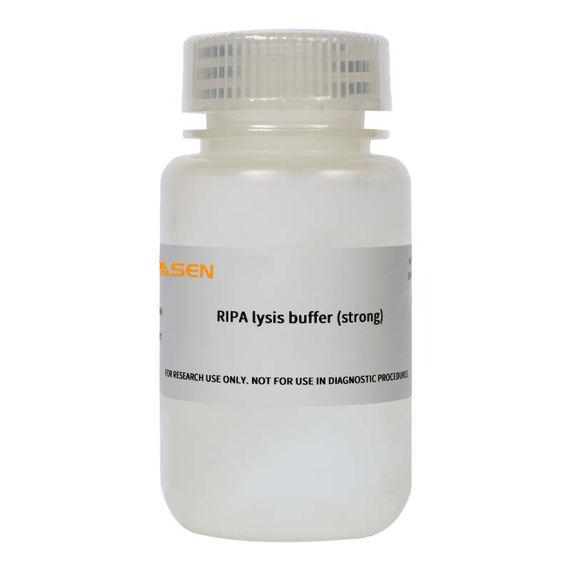
RIPA Lysis Buffer (Radio Immunoprecipitation Assay Buffer) is a traditional and widely used lysis solution for rapid disruption of cells and tissues. It effectively lyses cellular components from animal cells, including membrane, cytoplasmic, and nuclear fractions. Depending on the strength of the lysis buffer, RIPA buffers are generally categorized as strong, medium, or weak.
This product is a strong RIPA Lysis Buffer, containing multiple inhibitors such as sodium orthovanadate, sodium fluoride, EDTA, and leupeptin, which effectively prevent protein degradation. Protein samples obtained using this lysis buffer are suitable for routine applications such as Western blotting and immunoprecipitation (IP).
Storage
Ship with wet ice. This product should be stored at -20℃ for 1 year.Avoid repeated freeze-thaw cycles. Aliquotting is recommended for long-term storage.
Notes
1. PMSF must be provided by the user.
2. All steps involving sample lysis should be performed on ice or at 4°C.
3. Protein concentration of samples lysed with this RIPA buffer can be determined using a BCA Protein Assay Kit (Cat. No. 20201ES76). Due to the high concentration of detergents in this buffer, the Bradford method is not suitable for protein quantification of samples prepared with this lysis buffer.
4. For safety and hygiene, wear a lab coat and disposable gloves when handling reagents.
5. This product is intended for research use only.
Instructions
I. Cultured Cell Samples
1. Thaw the RIPA Lysis Buffer and mix thoroughly. Add PMSF to the appropriate volume of lysis buffer shortly before use (within a few minutes), to achieve a final PMSF concentration of 1 mM.
2. For adherent cells: Remove culture medium and wash cells once with PBS, physiological saline, or serum-free medium (washing is optional if serum proteins do not interfere). Add 150–250 μL of lysis buffer per well of a 6-well plate. Pipette up and down several times to ensure complete contact between the lysis buffer and cells. After complete lysis, no visible cell pellets should remain.
For suspension cells: Pellet cells by centrifugation and resuspend the pellet by tapping the tube firmly with your finger. Add 150–250 μL of lysis buffer per 6-well equivalent of cells. Tap the tube gently to ensure complete cell lysis. After complete lysis, no visible cell pellets should remain. If the cell number is high, divide the sample into aliquots of 0.5–1×10⁶ cells per tube before lysis.
3. After complete lysis, centrifuge at 10,000–14,000 g for 3–5 minutes. Collect the supernatant for downstream applications such as PAGE, Western blotting, or immunoprecipitation.
Note on Lysis Buffer Volume: Typically, 150 μL of lysis buffer per well of a 6-well plate is sufficient. However, if cell density is very high, the volume may be increased to 200 μL or 250 μL as needed.
II. Tissue Samples
1. Cut the tissue into small fragments.
2. Thaw the RIPA Lysis Buffer and mix thoroughly. Add PMSF shortly before use to achieve a final concentration of 1 mM.
3. Add lysis buffer at a ratio of 150–250 μL per 20 mg of tissue. (If lysis is insufficient, additional lysis buffer may be added. If higher protein concentration is desired, reduce the volume of lysis buffer accordingly.)
4. Homogenize the sample using a glass homogenizer until complete lysis is achieved.
5. After complete lysis, centrifuge at 10,000–14,000 g for 3–5 minutes. Collect the supernatant for downstream applications such as PAGE, Western blotting, or immunoprecipitation.
6. If the tissue sample is already very small, it may be cut into small pieces and directly lysed by adding lysis buffer and vigorous vortexing. After complete lysis, centrifuge and collect the supernatant for subsequent experiments. The advantage of direct lysis is convenience and no need for a homogenizer; the disadvantage is that lysis may be less complete compared to using a homogenizer.
[Note]: A small amount of transparent, gel-like material is often observed in the lysate when using RIPA buffer—this is a normal phenomenon and consists of complexes containing genomic DNA. If you are not analyzing proteins that bind tightly to genomic DNA, the supernatant can be directly collected by centrifugation for downstream applications. If you are analyzing proteins that bind tightly to DNA, sonication can be used to break up and disperse the gel-like material, followed by centrifugation to collect the supernatant. For the detection of common transcription factors such as NF-kappaB or p53, sonication is typically not required to achieve detectable results.
Documents:
Safety Data Sheet
20101_MSDS_HB250820_EN.PDF
Manuals
20101_Manual_Ver.EN20250820.pdf
Betaling en beveiliging
Uw betalingsinformatie wordt veilig verwerkt. We slaan geen creditcardgegevens op en hebben geen toegang tot uw creditcardinformatie.
Navraag
Misschien vind je het ook leuk
FAQ
Het product is alleen bedoeld voor onderzoeksdoeleinden en is niet bedoeld voor therapeutisch of diagnostisch gebruik bij mensen of dieren. Producten en inhoud worden beschermd door patenten, handelsmerken en auteursrechten die eigendom zijn van
Voor bepaalde toepassingen zijn mogelijk aanvullende intellectuele eigendomsrechten van derden vereist.

