


Frequently Bought Together
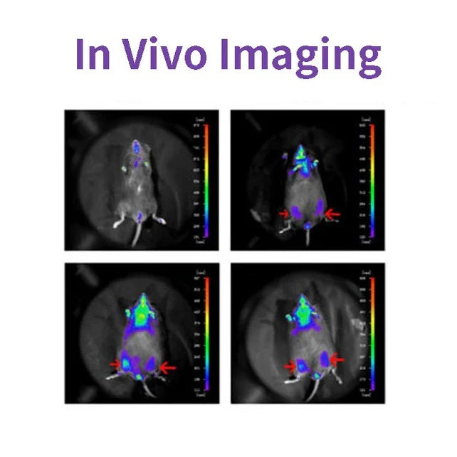
D-ルシフェリンはルシフェラーゼの一般的な基質であり、バイオテクノロジー、特に生体内イメージング技術で広く使用されています。その作用機序は、ルシフェリン(基質)がATPとルシフェラーゼの存在下で酸化されて光を発することです。フルオレセインが過剰の場合、生成される光の量子数はルシフェラーゼ濃度に等しく、正の相関関係があります(下の図を参照)。ルシフェラーゼをコードする遺伝子(Luc)を持つプラスミドが細胞にトランスフェクトされ、ラットやマウスなどの研究動物に導入されました。

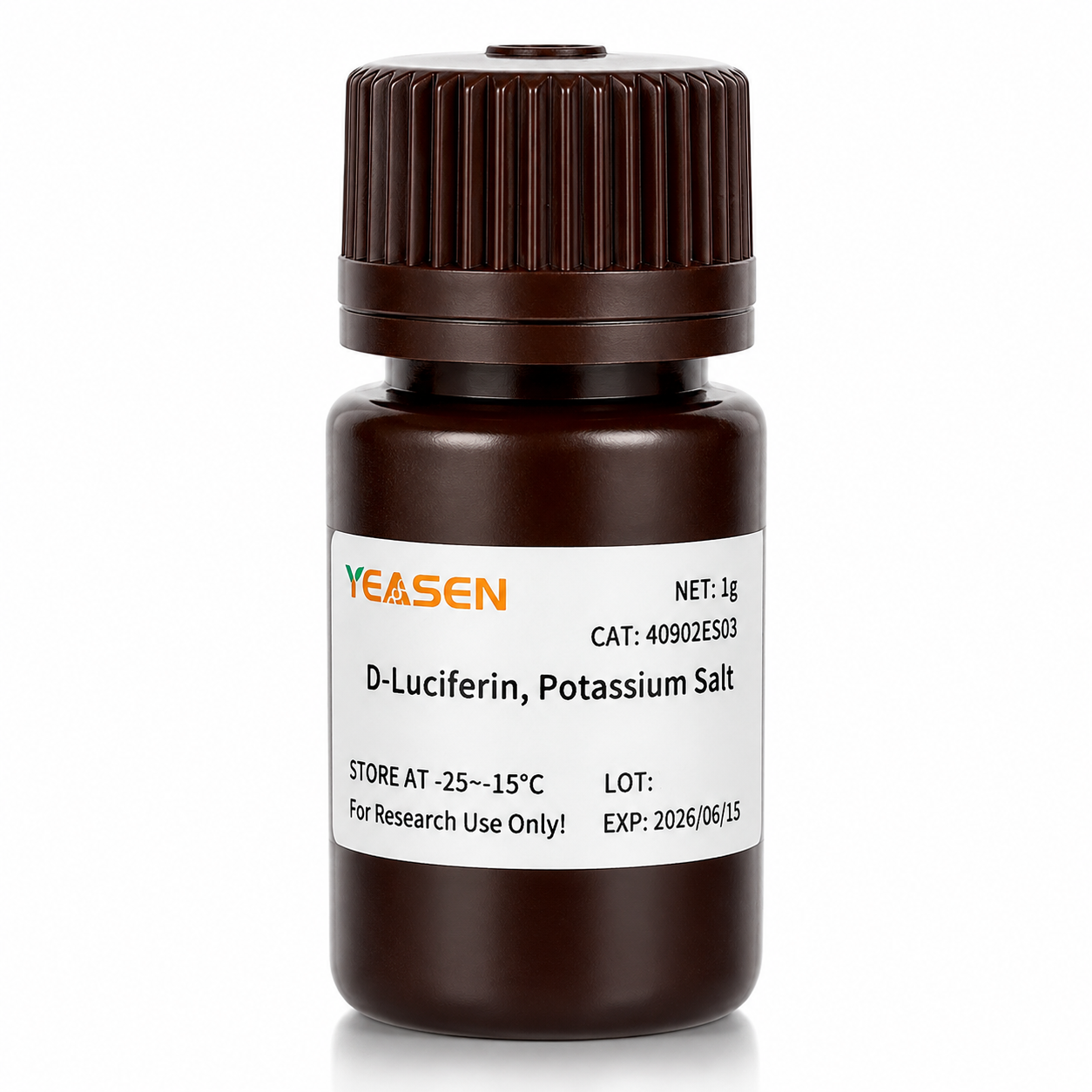
D-ルシフェリンは、ルシフェラーゼおよびATPレベル分析、レポーター遺伝子分析、ハイスループットシーケンシング、およびさまざまな汚染テストを含むin vitro研究でもよく使用されます。製品形態は、D-ルシフェリン(遊離酸)、D-ルシフェリン塩(ナトリウム塩およびカリウム塩)の3つです。主な違いは溶解性にあります。前者は水溶性で、後者は緩衝液系の溶解性です。低濃度のNaOHおよびKOH溶液などの弱塩基を除いて、これらは弱く溶けます。メタノールおよびDMSOに溶けます。後者は水または緩衝液に簡単に溶け、使いやすいです。
特徴
- 放射線は出ないので、生体にほとんど無害です。
- 生物発光、励起光源なし。
- 非常に感度が高いため、数百個の細胞でも検出できます。
- 浸透性は良好で、3〜4cmの組織深度も検出可能です。
- 高い信号対雑音比、強い蛍光信号、優れた耐干渉性。
アプリケーション
- ヌードマウスの腫瘍形成実験では、腫瘍剥離測定を行わず、浸潤なくリアルタイムで腫瘍の成長を観察しました。
- 腫瘍の成長や転移に対する投与の効果を試験するために、フルオレセイン基質は薬物実験に干渉することなく 3 時間以内に完全に除去できます。
- 動物における外来細胞の局在と分布が検出されました。
- 標的遺伝子または標的遺伝子のプロモーターをルシフェラーゼ遺伝子に融合して、薬物治療中または疾患進行中の遺伝子発現の変化を検出します。
- 幹細胞移植、生存、増殖を監視し、生体内での幹細胞の分布と移動を追跡します。
仕様
| 英語の同義語 | (S)-4,5-ジヒドロ-2-(6-ヒドロキシ-2-ベンゾチアゾリル)-4-チアゾールカルボン酸カリウム塩; D-ルシフェリンホタル、カリウム塩 |
| CAS番号 | 115144-35-9 |
| 式 | C11H7N2O3S2K |
| 分子量 | 318.42グラム/モル |
| 外観 | 淡黄色の粉末 |
| 溶解度 | 水に溶ける(60 mg/mL) |
コンポーネント
| コンポーネント番号 | 名前 | 40902ES01 | 40902ES02 | 40902ES03 | 40902ES08 |
| 40902 | D-ルシフェリン、カリウム塩 | 100mg | 500mg | 1グラム | 5g |
数字

「ホスホグリセリン酸ムターゼ1の新規アロステリック阻害剤が非小細胞肺癌の増殖と転移を抑制する」Cell Metab. 2019;30(6):1107-1119より引用
配送と保管
商品はドライアイスと一緒に発送され、-15℃~-25℃で1年間保存可能です。
[1] Gu Y、Wang Y、He L、et al。環状RNA circIPO11はヘッジホッグシグナルを介して肝臓がん誘発細胞の自己複製を促進する。Mol Cancer。2021;20(1):132。2021年10月14日発行。doi:10.1186/s12943-021-01435-2(IF:27.401)
[2] Huang K、Liang Q、Zhou Y、et al。ホスホグリセリン酸ムターゼ1の新規アロステリック阻害剤は非小細胞肺癌の増殖と転移を抑制する[Cell Metab. 2021年1月5日;33(1):223に掲載された訂正]。Cell Metab. 2019;30(6):1107-1119.e8. doi:10.1016/j.cmet.2019.09.014(IF:22.415)
[3] Dong X、Cheng R、Zhu S、et al。ヘテロ接合構造のWO<sub>2.9</sub>-WSe<sub>2</sub>ナノ放射線増感剤は、低線量での局所腫瘍アブレーションとチェックポイント遮断免疫療法の効果を高めます。ACS Nano。2020;14(5):5400-5416。doi:10.1021/acsnano.9b08962(IF:14.588)
[4] Zheng DW、Gao F、Cheng Q、et al。自然免疫を開始し、腫瘍免疫療法を改善するためのワクチンベースのナノシステム。Nat Commun。2020;11(1):1985。2020年4月24日発行。doi:10.1038 / s41467-020-15927-0(IF:12.121)
[5] Hu J、Su H、Cao H、et al。AUXIN RESPONSE FACTOR7は、DELLAとAUX/IAAタンパク質間の相互作用を介してジベレリンとオーキシンのシグナル伝達を統合し、ポプラの形成層の活動を制御します。Plant Cell。2022;34(7):2688-2707。doi:10.1093/plcell/koac107(IF:11.277)
[6] Qi P、Huang M、Hu X、他。 Ralstonia solanacearum エフェクターは、TGA 転写因子を標的にしてサリチル酸シグナル伝達を破壊します。植物細胞。 2022;34(5):1666-1683。土井:10.1093/plcell/koac015(IF:11.277)
[7] Huang X、Qiu M、Wang T、et al。腹腔内播種性卵巣癌治療のためのキャリアフリー多機能ナノ医薬品。J Nanobiotechnology。2022;20(1):93。2022年2月22日発行。doi:10.1186/s12951-022-01300-4(IF:10.435)
[8] Tang Y、Lin S、Yin S、et al. マグネシウム粒子の分解に基づくその場ガス発泡:注入可能なマクロポーラスハイドロゲルを製造するための新しいアプローチ。Biomaterials. 2020;232:119727. doi:10.1016/j.biomaterials.2019.119727(IF:10.273)
[9] Zhang X、Zheng S、Hu C、et al。がん関連線維芽細胞誘導lncRNA UPK1A-AS1は、効率的な二本鎖切断修復を介して膵臓がんにプラチナ耐性を付与します。Oncogene。2022;41(16):2372-2389。doi:10.1038/s41388-022-02253-6(IF:9.867)
[10] Qiao K, Liu Y, Xu Z, et al. RNA m6Aメチル化はHippo経路を介して肝細胞癌における血管形成模倣の形成を促進する。血管新生。2021;24(1):83-96. doi:10.1007/s10456-020-09744-8(IF:9.780)
[11] Wu Q、Kuang K、Lyu M、et al. 植物の発育の光制御におけるマイクロタンパク質によるPIFとEIN3のアロステリック不活性化。Proc Natl Acad Sci US A. 2020;117(31):18858-18868. doi:10.1073/pnas.2002313117(IF:9.412)
[12] Meng DF、Sun R、Liu GY、et al。S100A14はIRAK1の分解を介してNF-κBシグナル伝達を阻害することにより鼻咽頭癌の転移を抑制する。Oncogene。2020;39(30):5307-5322。doi:10.1038/s41388-020-1363-8(IF:7.971)
[13] Gao Y、Wang J、Han H、et al。乳がん切除後の再発を抑えるためのナノ粒子含有ポリカプロラクトンインプラント。Nanoscale。2021;13(34):14417-14425。2021年9月2日発行。doi:10.1039/d1nr04125h(IF:7.790)
[14] Jiang W、Li T、Guo J、et al。二重特異性c-Met/PD-L1 CAR-T細胞は肝細胞癌に対する治療効果を高める。Front Oncol. 2021;11:546586。2021年3月10日発行。doi:10.3389/fonc.2021.546586(IF:6.244)
[15] Zhou Y、Zhou C、Zou Y、et al. ドキソルビシンとクルクミンを効率的に共送達し、相乗的に腫瘍転移を抑制するためのマルチpH感受性ポリマー薬物複合体混合ミセル。Biomater Sci. 2020;8(18):5029-5046. doi:10.1039/d0bm00840k(IF:6.183)
[16] Zhao X、Liu X、Zhang P、et al。同所性肝細胞癌の治療のための腹腔内トリプトライドデポとしての注射用ペプチドハイドロゲル。Acta Pharm Sin B。2019;9(5):1050-1060。doi:10.1016/j.apsb.2019.06.001(IF:5.808)
[17] Li M、Wang J、Yu Y、et al。ビスフォスフォネート関連顎骨壊死患者の歯肉から得られた間葉系幹細胞の特性。Stem Cell Rev Rep. 2022;18(1):378-394. doi:10.1007/s12015-021-10241-8(IF:5.739)
[18] Zhang X、Li Y、Ji J、et al。Gadd45gは胚性幹細胞の分化を開始し、乳がん細胞の発がんを抑制する。Cell Death Discov。2021;7(1):271。2021年10月2日発行。doi:10.1038/s41420-021-00667-x(IF:5.241)
[19] Wu D, Lv J, Zhao R, et al. PSCAは胃癌におけるキメラ抗原受容体T細胞の標的である。Biomark Res. 2020;8:3. 2020年1月28日発行。doi:10.1186/s40364-020-0183-x(IF:4.866)
[20] Qiao K, Chen C, Liu H, Qin Y, Liu H. Pininはm6A修飾を制御することで肝細胞癌の上皮間葉転換を誘導する。J Oncol. 2021;2021:7529164。2021年12月7日発行。doi:10.1155/2021/7529164(IF:4.375)
[21] Li K、Liu T、Chen J、Ni H、Li W。乳がん由来エクソソーム中のSurvivinはSOD1をアップレギュレーションすることで線維芽細胞を活性化し、そのフィードバックががんの増殖と転移を促進する。J Biol Chem. 2020;295(40):13737-13752. doi:10.1074/jbc.RA120.013805(IF:4.238)
[22] Kong Y、Feng Z、Chen A、et al。天然フラボノイドガランギンはグリオーブラストーマでアポトーシス、ピロプトーシス、オートファジーを誘発する。Front Oncol. 2019;9:942。2019年9月27日発行。doi:10.3389/fonc.2019.00942(IF:4.137)
[23] Kong Y、Feng Z、Chen A、et al。天然フラボノイドガランギンはグリオーブラストーマでアポトーシス、ピロプトーシス、オートファジーを誘発する。Front Oncol. 2019;9:942。2019年9月27日発行。doi:10.3389/fonc.2019.00942(IF:4.137)
[24] Hu J、Wang Y、Yuan Y. APE1酸化還元機能阻害剤は、in vitroおよびin vivoでγヘルペスウイルスの複製を効果的に阻害する。Antiviral Res. 2021;185:104985. doi:10.1016/j.antiviral.2020.104985(IF:4.101)
[25] Li Q, Wei D, Feng F, et al. α2,6結合シアリン酸はニューカッスル病ウイルスを用いた癌腫瘍溶解性ウイルス療法の高親和性受容体として機能する。J Cancer Res Clin Oncol. 2017;143(11):2171-2181. doi:10.1007/s00432-017-2470-y(IF:3.503)
[26] Han B, Jiang P, Liu W, et al. 乳がんにおけるダウコステロールリノール酸の役割:アポトーシスと転移に関する研究。J Agric Food Chem. 2018;66(24):6031-6041. doi:10.1021/acs.jafc.8b01387(IF:3.412)
[27] Hu H、Zhang Z、Wang R、et al。iRFP720を安定発現するBGC823細胞株は、有望な蛍光イメージング能力を備え、その主要な特性を保持しています。DNA Cell Biol。2020;39(5):900-908。doi:10.1089/dna.2019.5057(IF:3.314)
[28] Ni XR、Zhao YY、Cai HP、et al。トランスフェリン受容体1を標的とした光学イメージングによるマウスモデルにおける神経膠腫縁の特定。J Neurooncol. 2020;148(2):245-258. doi:10.1007/s11060-020-03527-3(IF:3.267)
[29] Feng H, Tang J, Zhang P, Miao Y, Wu T, Cheng Z. Ilex cornutaの葉からの抗脂肪生成18,19-セコウルサン立体異性体とオレアン型サポニン。植物化学。2020;175:112363. doi:10.1016/j.phytochem.2020.112363(IF:3.044)
[30] Zhao Y, Li T, Tian S, et al. EIF4A1を標的としたMYC増幅グループ3髄芽腫の効果的な阻害。Cancer Manag Res. 2020;12:12473-12485。2020年12月3日発行。doi:10.2147/CMAR.S278844(IF:2.886)
Storage
FAQ
1) Solubility: The salt form is easily soluble in water. The solubility of potassium salt is 60mg/ml, and that of sodium salt is 100mg/ml. Free acid is not easily soluble in water. It can be dissolved by a weak alkaline solution of sodium bicarbonate, and its solubility in methanol is 10mg/ml, and in DMSO is 50mg/ml.
2) Toxicity: The salt form is more convenient to use during the process, especially in in vivo imaging experiments, as it can dissolve in water and the reaction toxicity will be smaller (Fluorescein is a low-molecular-weight organic compound composed of phenylthiazole and thiazole carboxyl group, with low toxicity).
3) Usage effect: There is no significant difference. In in vivo experimental studies, the usage rate of potassium salt is higher.
2. Native coelenterazine is an action substrate for various luciferases such as sea kidney luciferase (Rluc) and Gaussia luciferase (Gluc).
1. Instruments with a biochemiluminescence detection module. The light produced by luciferin can be detected by a photometer or a scintillation counter. Common in vivo imaging instruments: such as the IVIS® Lumina small animal in vivo imaging system, the In-Vivo Xtreme multi-mode small animal in vivo imaging instrument from Bruker, Germany.
2. Multi-functional microplate readers: It is necessary to confirm with the instrument manufacturer whether it is a specific biochemiluminescence detection module (note: cannot use a fluorescence microscope).
2. The cells themselves also contain a certain amount of ATP and magnesium, which can promote the reaction. In most cases, the ATP and magnesium contained in the cells are sufficient for the reaction of D-fluorescein salt, but the content varies depending on the type of cells. You can first try using a culture medium without additional ATP and magnesium, and conduct the experiment normally to see the results.
1) Injection method: It can be done through intraperitoneal injection or tail vein injection.
2) Injection volume: The scientific approach is to evaluate the injection dose based on the kinetic curve. The initial attempted injection dose is: 150mg substrate/kg of mouse body weight. Therefore, the purchase quantity can be calculated according to the above method: If there are 10 mice, 22 - 25g, then 33 - 37.5mg of substrate is required.
Citations
Documents
| Brochure |
|---|
| In_vivo_imaging_Leaving_Tumor_Cells_Nowhere_to_Hide.pdf |


あなたも好きかもしれません
問い合わせ
よくある質問
この製品は研究目的のみに使用され、人間や動物の治療や診断に使用することを意図したものではありません。製品とコンテンツは、
特定のアプリケーションでは、追加のサードパーティの知的財産権が必要になる場合があります。