
Keterangan
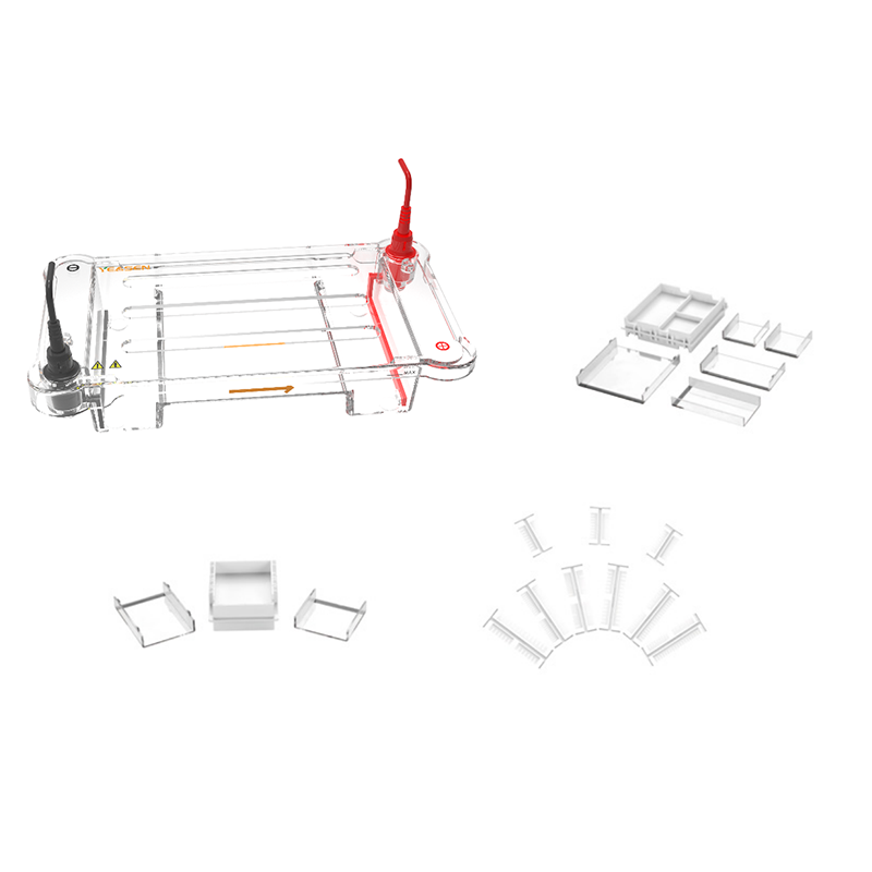
Horizontal electrophoresis equipped with the function of adding sample by pipette is mainly used for electrophoresis of the agarose gel of DNA and RNA, whose special-used gel casting bench and flexible combination of tray with ear-shaped structure are convenient to be used. It can conduct the experiment of 96-hole PCR sample electrophoresis with different volume of agarose and size like 6.5×6.5 cm, 6.5×13 cm, 13×6.5 cm, 13×13 cm etc. The instrument mainly consists of gel tray, lower body, upper body, gel-making kit, comb etc.
Features
High-throughput: 13×13 cm in size, capable of handling 108 samples (including markers) at one time, specially designed for multi-sample analysis.
Convenient operation: special glue filling table, free combination of pallets of various specifications; The 14-,19-, and 27-tooth combs all support 8-channel and 12-tooth guns.
High temperature resistance: no need to dry the agarose until warm and then pour the gel, and it will not be deformed at a high temperature of 100°C.
Acid and alkali resistance: the tank body is integrated and molded, so there is no need to worry about long-term soaking and leakage.
Good conductivity: bold conductive platinum wire, better conductivity.
Applications
DNA fragment isolation and identification
Nucleic acid quality testing
Cloning and genetic engineering
Genetic Analysis and Forensics
Research and teaching experiments
Specifications
|
Specification Category |
Details |
|
Main Tank Dimensions |
300(L) × 155(W) × 100(H) mm |
|
Tray Area (W×L) |
Standard: |
|
Combs |
|
|
Simultaneous Gel Casting Capacity |
1-4 gels |
|
Buffer Capacity |
1000 mL |
|
Maximum Voltage |
200 V |
|
Maximum Power |
40 W |
Components
|
Components No. |
Name |
80230ES01 |
|
80230 |
horizontal electrophoresis |
1 unit |
Shipping and Storage
The product can be stored at room temperature for five year.
Operation instructions
1. Put the gel-making frame on the horizontal desk and put the gel tray into the grid of gel-making frame accordingly then put the comb in the narrow slot.4 types of gel can be made in the gel-making frame like 13×13 cm, 13×6.5 cm, 6.5×13 cm, 6.5×6.5 cm according to the actual needs.
2. Make the agarose solution with proper concentration by electrophoresis buffer according to the size of separated DNA fragment: measure the dry powder of agarose accurately and put it in the conical flask or the glass bottle with fixed volume of electrophoresis buffer, and then use the glass stick to stir it evenly and put it into the boiling water or microwave oven for being heated until the agarose is fused (to determine the concentration of agarose according to the attached list).
3. Put the gel into the gel tray slowly while it is slightly cooled, the ideal thickness of gel is 3~5mm (Note: avoid the bubble in the gel).
4. Let the gel coagulates completely for 30~45min in room temperature (The coagulation period can also be shortened by putting it in the 4℃ refrigerator after the gel coagulates slightly). Take out the comb carefully and put gel in the cell, the side of hole is close to cathode (black end).
5. Put the buffer in the electrophoresis cell and keep the surface of buffer at least 2mm higher than the gel (Note: TAE buffer should be replaced after 2 to 3 times, the TBE buffer can be used for around 10 times).
Mix proper amount of DNA sample and 10×buffer (analyze single DNA sample, such as L bacteriophage or plasmid DNA, each sample-adding hole with width of 5mm is suitable for 100~500ng DNA. The resolution is not decreased obviously when 20~30μg is added if sample consists of many DNA fragments, such as DNA enzyme digestion sample of mammal). Use the pipette to add the sample with proper amount of standard DNA molecular weight into the right side hole and left side hole.
Lid the electrophoresis cell after sample adding and power on by 5~8V/cm, the distance in which should be matched with the measured distance between anode and cathode. The bubble is created by electrolytic action. DNA migrates to the anode (red plug).The period of electrophoresis is determined by the length of gel voltage, and the size of DNA fragment. The longer the gel is, the lower the voltage is, the bigger the DNA fragment is, the more time required. However the resolution of big DNA fragment is very low and the band is not clear if the high voltage is adopted (The voltage per centimeter of gel is less than 8V because the high voltage causes the lower resolution. The electrophoresis migration rate of linear DNA molecular is increased as voltage rises accordingly only in the lower voltage.).
When indicator migrates to the bottom of gel, power off and take out sample and put it in the EB solution for being dyed for 5~10min (EB will be resolved in the sunshine and should be stored in the dark room),Observe the sample in UV Transilluminator and take photo if necessary (EB can be put in the gel during the gel-making process).
Documents:
Safety Data Sheet
Manuals
Pembayaran & Keamanan
Informasi pembayaran Anda diproses dengan aman. Kami tidak menyimpan detail kartu kredit atau memiliki akses ke informasi kartu kredit Anda.
Anda mungkin juga menyukai
Pertanyaan
FAQ
Produk ini hanya untuk keperluan penelitian dan tidak ditujukan untuk penggunaan terapeutik atau diagnostik pada manusia atau hewan. Produk dan konten dilindungi oleh paten, merek dagang, dan hak cipta milik
Aplikasi tertentu mungkin memerlukan hak kekayaan intelektual pihak ketiga tambahan.