Description
CHO Host Cell DNA Residue Detection Kit is used for the quantitative analysis of CHO host cell DNA residuce in intermediate samples, semi-finished and finished products of various biological products.
This kit adopts Taqman fluorescent probe and the polymerase chain reaction (PCR) method, which has fg level minimum detection limit and can specifically and quickly detect the residual CHO cell DNA. The kit needs to be used together with the the Residual DNA Sample Preparation Kit (Cat# 18461ES or 18469).
Features
High anti-interference capability – In complex sample matrices, spike recovery rates are consistently between 70% and 130%.
High sensitivity – Limit of quantification (LLOQ) as low as 10 fg/μL.
Excellent precision – Intra-assay CV <10%; inter-assay (intermediate) CV <15%.
Strong specificity – Specifically detects HEK 293 residual DNA without interference from other genomic DNA.
Robust contamination control – Includes UDG enzyme to digest potential carryover from PCR products and aerosols, preventing non-specific amplification.
Comprehensive validation – Fully validated according to ICH Q2 (R2) guidelines; complete validation reports available.
Regulatory compliance – Verified according to EP 2.6.7, JP G3, and USP <63> pharmacopeia standards.
Quality assurance – All enzymes are self-developed and industrialized, ensuring stable supply and consistent kit performance.
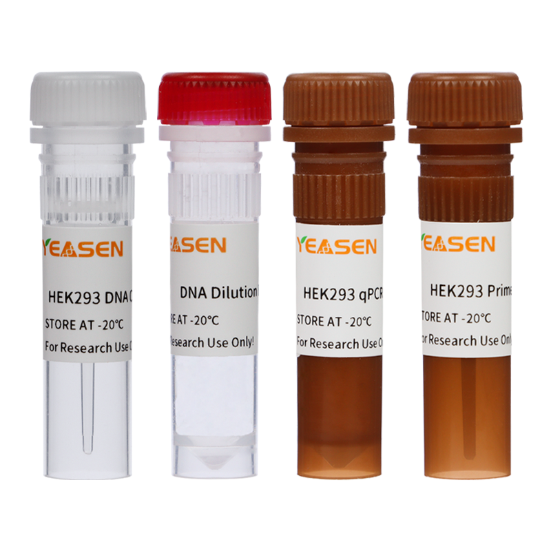
Components
|
Components No. |
Name |
41331ES50 (50 T) |
41331ES60 (100 T) |
|
A |
HEK293 qPCR Mix (UDG plus) |
0.75 mL |
1.5 mL |
|
B |
HEK293 Primer&Probe Mix |
250 μL |
500 μL |
|
C |
DNA Dilution Buffer |
2×1.8 mL |
4×1.8 mL |
|
D |
HEK293 DNA Control(30 ng/μL) |
25 μL |
50 μL |
Specifications
|
Linearity |
Standard curve range: 30 fg/μL – 300 pg/μL Correlation coefficient (R²): ≥ 0.98 Amplification efficiency: 90%–110% |
|
Accuracy |
Recovery rate: 50%–150% (industry standard 70%–130%) |
|
Precision |
Repeatability: CV < 15% Intermediate precision: CV < 15% |
|
Sensitivity |
Limit of quantification (LOQ): 10 fg/μL |
|
Specificity |
No interference from other genomic DNA sources |
|
Stability |
Accelerated stability: 14 days at 37 °C Freeze–thaw stability: ≥ 10 cycles |
|
Blank Control |
No-template control (NTC): In 96 repeated qPCR runs, all Ct values ≥ 35 or no amplification observed |
|
Applicable instrument models |
Bio-Rad: CFX96 Optic Module. Thermo Scientific: ABI 7500; ABI Quant Studio 5. SLAN-96S. |
|
Application |
Detection of residual HEK293 host cell DNA in viral and cell products, including lentivirus and AAV production, as well as other HEK293-derived bioproducts. |
Storage
This product should be stored at -25~-15℃ for 2 years.
Both 41331-A and 41331-B should be stored protected from light.
Figures
1. Good Linearity

Figure 1. Standard curve (left) and amplification curve plot (right) for HEK293 DNA (3G).
The linear range of the HEK293 Host Cell Residual DNA Detection Kit (3G) is 30 fg/μL to 300 pg/μL, with an R² value of 1.0 and an amplification efficiency of 100.45%. The coefficient of variation (CV) for detection values across concentrations is <15%.
2. Sensitivity: Limit of quantification (LOQ) - 0.3 fg/μL

Figure 2. qPCR detection of HEK293 DNA at 10 fg/μL.
HEK293 DNA (3G) at the lowest standard curve concentration (3 fg/μL) and below was tested with 10 replicates per concentration. Results showed that at concentrations ≥10 fg/μL, the CV was <20%, indicating that the limit of quantification (LOQ) of the HEK293 Host Cell DNA Residual Detection Kit (3G) is 10 fg/μL.
3. High Recovery rate: 50%–150%
Table 1. Recovery of HEK293 DNA in Blank Samples

Spike recovery rates for all concentrations tested were between 70% and 130%, with coefficients of variation (CV) below 20%, demonstrating consistent performance of the assay across a wide dynamic range.
Table 2. HEK293 DNA Recovery in Various Sample Matrices

HEK293 DNA (3 pg/μL) was spiked into five different background matrices (high protein, high nucleic acid, high salt, high pH, low pH) and extracted using the same procedure. Recovery rates ranged from 70% to 130%, with CVs below 20% for all matrices, demonstrating robust performance across diverse sample backgrounds.
Documents
Safety Data Sheet
Manuals
41331_Manual_Ver.EN20240517.pdf
Related Blog
Host Cell Residual DNA Detection Kits facilitate Biologics R&D and Manufacturing
Advancing Biopharmaceutical Quality Control: Unveiling the Science of HEK293 Residual Detection
Payment & Security
Your payment information is processed securely. We do not store credit card details nor have access to your credit card information.
Inquiry
You may also like
FAQ
The product is for research purposes only and is not intended for therapeutic or diagnostic use in humans or animals. Products and content are protected by patents, trademarks, and copyrights owned by Yeasen Biotechnology. Trademark symbols indicate the country of origin, not necessarily registration in all regions.
Certain applications may require additional third-party intellectual property rights.
Yeasen is dedicated to ethical science, believing our research should address critical questions while ensuring safety and ethical standards.


