


Frequently Bought Together
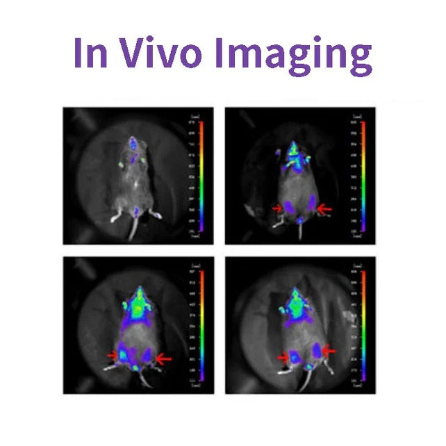
D-루시페린은 루시페라아제의 일반적인 기질이며, 생명공학, 특히 생체 내 영상 기술 전반에 널리 사용됩니다. 작용 기전은 루시페린(기질)이 ATP와 루시페라아제가 있는 상태에서 산화되어 빛을 방출할 수 있다는 것입니다. 플루오레세인이 과도할 때 생성되는 빛의 양자 수는 루시페라아제 농도와 양의 상관 관계가 있습니다(아래 그림 참조). 루시페라아제 인코딩 유전자(Luc)를 운반하는 플라스미드를 세포에 형질 도입하여 쥐와 생쥐와 같은 연구 동물에 도입했습니다.

D-루시페린은 루시페라아제 및 ATP 수치 분석, 리포터 유전자 분석, 고처리량 시퀀싱 및 다양한 오염 검사를 포함한 시험관 내 연구에도 일반적으로 사용됩니다. D-루시페린(유리산), D-루시페린 염(나트륨 염 및 칼륨 염)의 세 가지 제품 형태가 있습니다. 주요 차이점은 용해도에 있습니다. 전자의 수용성과 완충액 시스템의 용해성입니다. 이들은 낮은 농도의 NaOH 및 KOH 용액과 같은 약염기에서 제외하고는 약하게 용해됩니다. 메탄올 및 DMSO에 용해됩니다. 후자는 물이나 완충액에 쉽게 용해되어 사용하기 쉽습니다.
특징
- 방사선이 없으므로 생물체에 거의 무해합니다.
- 생물발광, 여기 광원 없음.
- 너무 민감해서 수백 개의 세포에서도 감지할 수 있어요.
- 침투성이 양호하여 3-4cm 조직 깊이도 감지할 수 있습니다.
- 높은 신호대잡음비, 강력한 형광신호, 우수한 간섭방지 기능을 갖추고 있습니다.
응용 프로그램
- 누드 마우스를 대상으로 한 종양 형성 실험에서는 종양 제거 측정 없이 실시간으로 침습 없이 종양 성장을 관찰했습니다.
- 종양 성장이나 전이에 대한 투여 효과를 시험하기 위해, 형광물질 기질은 약물 실험에 방해가 되지 않고 3시간 이내에 완전히 제거될 수 있습니다.
- 동물에서 이물질 세포의 국소화와 분포가 감지되었습니다.
- 표적 유전자 또는 표적 유전자의 프로모터는 루시퍼라제 유전자에 융합되어 약물 치료 또는 질병 진행 중에 유전자 발현의 변화를 감지합니다.
- 줄기세포 이식, 생존 및 증식을 모니터링합니다. 생체 내에서 줄기세포의 분포와 이동을 추적합니다.
명세서
| 영어 동의어 | (S)-4,5-디하이드로-2-(6-하이드록시-2-벤조티아졸릴)-4-티아졸카르복실산 칼륨염; D-루시페린 반딧불이, 칼륨염 |
| CAS 번호 | 115144-35-9 |
| 공식 | C11H7N2O3S2K |
| 분자량 | 318.42g/몰 |
| 모습 | 연한 노란색 분말 |
| 용해도 | 물에 용해됨(60 mg/mL) |
구성 요소
| 구성품 번호 | 이름 | 40902ES01 | 40902ES02 | 40902ES03 | 40902ES08 |
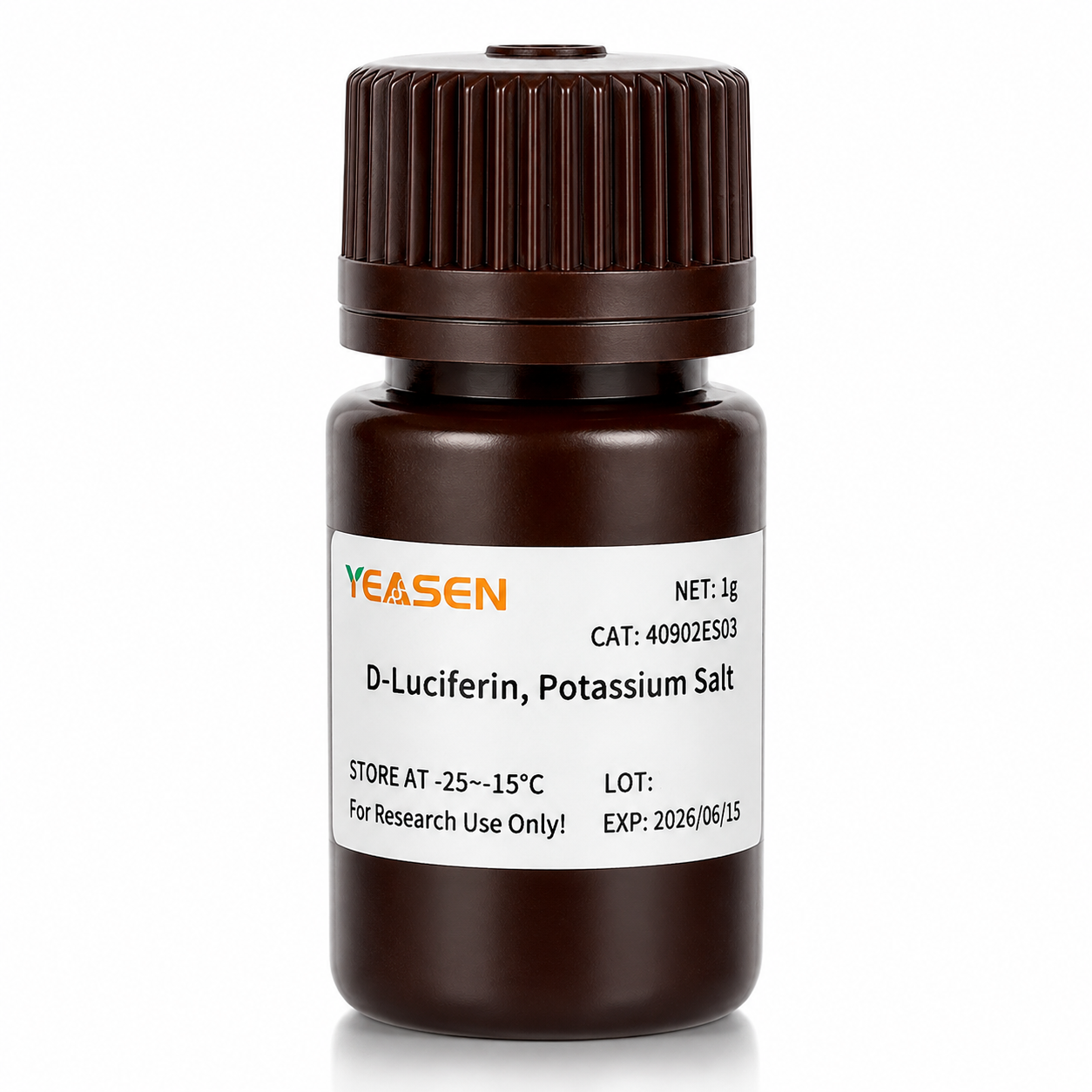
| 40902 | D-루시페린, 칼륨염 | 100mg | 500mg | 1g | 5g |
그림

"Phosphoglycerate Mutase 1의 새로운 알로스테릭 억제제는 비소세포 폐암의 성장 및 전이를 억제합니다, Cell Metab. 2019;30(6):1107-1119에서 인용
배송 및 보관
본 제품은 건조아이스와 함께 배송되며 -15℃ ~ -25℃에서 1년간 보관이 가능합니다.
[1] Gu Y, Wang Y, He L, et al. 원형 RNA circIPO11은 Hedgehog 신호를 통해 간암 개시 세포의 자가 재생을 촉진합니다. Mol Cancer. 2021;20(1):132. 2021년 10월 14일 출판. doi:10.1186/s12943-021-01435-2(IF:27.401)
[2] Huang K, Liang Q, Zhou Y, et al. Phosphoglycerate Mutase 1의 새로운 알로스테릭 억제제는 비소세포 폐암의 성장 및 전이를 억제합니다 [게시된 수정 사항은 Cell Metab. 2021 Jan 5;33(1):223에 나타남]. Cell Metab. 2019;30(6):1107-1119.e8. doi:10.1016/j.cmet.2019.09.014(IF:22.415)
[3] Dong X, Cheng R, Zhu S, et al. 이종 접합 구조의 WO<sub>2.9</sub>-WSe<sub>2</sub> 나노방사선감각제는 저방사선량에서 국소 종양 소거 및 체크포인트 차단 면역요법을 증가시킵니다. ACS Nano. 2020;14(5):5400-5416. doi:10.1021/acsnano.9b08962(IF:14.588)
[4] Zheng DW, Gao F, Cheng Q, et al. 선천 면역 개시 및 종양 면역 요법 개선을 위한 백신 기반 나노시스템. Nat Commun. 2020;11(1):1985. 2020년 4월 24일 출판. doi:10.1038/s41467-020-15927-0(IF:12.121)
[5] Hu J, Su H, Cao H, et al. AUXIN RESPONSE FACTOR7은 DELLA와 AUX/IAA 단백질 간의 상호 작용을 통해 지베렐린과 오신 신호를 통합하여 포플러의 속씨 활동을 조절합니다. Plant Cell. 2022;34(7):2688-2707. doi:10.1093/plcell/koac107(IF:11.277)
[6] Qi P, Huang M, Hu X 등. Ralstonia solanacearum effector는 TGA 전사 인자를 표적으로 삼아 살리실산 신호 전달을 파괴합니다. 식물세포. 2022;34(5):1666-1683. doi:10.1093/plcell/koac015(IF:11.277)
[7] Huang X, Qiu M, Wang T, et al. 복강내 확산성 난소암 치료를 위한 캐리어 없는 다기능 나노의학. J Nanobiotechnology. 2022;20(1):93. 2022년 2월 22일 출판. doi:10.1186/s12951-022-01300-4(IF:10.435)
[8] Tang Y, Lin S, Yin S, et al. 마그네슘 입자 분해를 기반으로 한 현장 가스 발포: 주입 가능한 거대 다공성 하이드로젤을 제조하는 새로운 접근법. 생체재료. 2020;232:119727. doi:10.1016/j.biomaterials.2019.119727(IF:10.273)
[9] Zhang X, Zheng S, Hu C, et al. 암 관련 섬유아세포 유도 lncRNA UPK1A-AS1은 효율적인 이중 가닥 절단 복구를 통해 췌장암에서 백금 저항성을 부여합니다. Oncogene. 2022;41(16):2372-2389. doi:10.1038/s41388-022-02253-6(IF:9.867)
[10] Qiao K, Liu Y, Xu Z, et al. RNA m6A 메틸화는 히포 경로를 통해 간세포암에서 혈관신생 모방 형성을 촉진합니다. 혈관신생. 2021;24(1):83-96. doi:10.1007/s10456-020-09744-8(IF:9.780)
[11] Wu Q, Kuang K, Lyu M, et al. 식물 발달의 빛 제어에서 미세단백질에 의한 PIF 및 EIN3의 이성질 비활성화. Proc Natl Acad Sci US A. 2020;117(31):18858-18868. doi:10.1073/pnas.2002313117(IF:9.412)
[12] Meng DF, Sun R, Liu GY, et al. S100A14는 IRAK1 분해를 통해 NF-kB 신호 전달을 억제하여 비인두암 전이를 억제합니다. Oncogene. 2020;39(30):5307-5322. doi:10.1038/s41388-020-1363-8(IF:7.971)
[13] Gao Y, Wang J, Han H, et al. 절제 후 유방암 재발에 대항하기 위한 나노입자 함유 폴리카프로락톤 임플란트. 나노스케일. 2021;13(34):14417-14425. 2021년 9월 2일 출판. doi:10.1039/d1nr04125h(IF:7.790)
[14] Jiang W, Li T, Guo J, et al. 이중특이성 c-Met/PD-L1 CAR-T 세포는 간세포암에 대한 치료 효과를 강화했습니다. Front Oncol. 2021;11:546586. 2021년 3월 10일 출판. doi:10.3389/fonc.2021.546586(IF:6.244)
[15] Zhou Y, Zhou C, Zou Y 등. 종양 전이를 시너지적으로 억제하기 위한 도코루비신과 쿠르쿠민의 효율적인 공동 전달을 위한 다중 pH 민감성 폴리머-약물 접합체 혼합 미셀. Biomater Sci. 2020;8(18):5029-5046. doi:10.1039/d0bm00840k(IF:6.183)
[16] Zhao X, Liu X, Zhang P, et al. 정형외과 간세포암 치료를 위한 복강내 트립톨리드 저장소로서의 주입형 펩타이드 하이드로젤. Acta Pharm Sin B. 2019;9(5):1050-1060. doi:10.1016/j.apsb.2019.06.001(IF:5.808)
[17] Li M, Wang J, Yu Y, et al. 턱뼈 환자의 치은 비스포스포네이트 관련 골괴사에서 유래한 중간엽 줄기세포의 특성화. Stem Cell Rev Rep. 2022;18(1):378-394. doi:10.1007/s12015-021-10241-8(IF:5.739)
[18] Zhang X, Li Y, Ji J, et al. Gadd45g는 배아줄기세포 분화를 개시하고 유방세포 발암을 억제합니다. Cell Death Discov. 2021;7(1):271. 2021년 10월 2일 출판. doi:10.1038/s41420-021-00667-x(IF:5.241)
[19] Wu D, Lv J, Zhao R, et al. PSCA는 위암에서 키메라 항원 수용체 T 세포의 표적입니다. Biomark Res. 2020;8:3. 2020년 1월 28일 출판. doi:10.1186/s40364-020-0183-x(IF:4.866)
[20] Qiao K, Chen C, Liu H, Qin Y, Liu H. Pinin은 m6A 변형을 조절하여 간세포암에서 상피세포-중간엽세포 전환을 유도합니다. J Oncol. 2021;2021:7529164. 2021년 12월 7일 출판. doi:10.1155/2021/7529164(IF:4.375)
[21] Li K, Liu T, Chen J, Ni H, Li W. 유방암 유래 엑소좀의 Survivin은 SOD1을 상향 조절하여 섬유아세포를 활성화하고, 이 피드백은 암 증식과 전이를 촉진합니다. J Biol Chem. 2020;295(40):13737-13752. doi:10.1074/jbc.RA120.013805(IF:4.238)
[22] Kong Y, Feng Z, Chen A, et al. 천연 플라보노이드 갈란긴은 신경교종에서 세포사멸, 피로프토시스 및 자가포식을 유발합니다. Front Oncol. 2019;9:942. 2019년 9월 27일 출판. doi:10.3389/fonc.2019.00942(IF:4.137)
[23] Kong Y, Feng Z, Chen A, et al. 천연 플라보노이드 갈란긴은 신경교종에서 세포사멸, 피로프토시스 및 자가포식을 유발합니다. Front Oncol. 2019;9:942. 2019년 9월 27일 출판. doi:10.3389/fonc.2019.00942(IF:4.137)
[24] Hu J, Wang Y, Yuan Y. APE1 산화환원 기능 억제제는 시험관 내 및 생체 내에서 γ-헤르페스바이러스 복제를 효과적으로 억제합니다. 항바이러스 연구. 2021;185:104985. doi:10.1016/j.antiviral.2020.104985(IF:4.101)
[25] Li Q, Wei D, Feng F, et al. α2,6-연결된 시알산은 뉴캐슬병 바이러스를 이용한 항암용해성 바이러스 요법을 위한 고친화성 수용체 역할을 합니다. J Cancer Res Clin Oncol. 2017;143(11):2171-2181. doi:10.1007/s00432-017-2470-y(IF:3.503)
[26] Han B, Jiang P, Liu W, et al. 유방암에 대한 Daucosterol Linoleate의 역할: 세포사멸 및 전이에 관한 연구. J Agric Food Chem. 2018;66(24):6031-6041. doi:10.1021/acs.jafc.8b01387(IF:3.412)
[27] Hu H, Zhang Z, Wang R, et al. iRFP720의 안정적인 발현을 갖는 BGC823 세포주는 유망한 형광 이미징 능력과 함께 기본 특성을 유지합니다. DNA Cell Biol. 2020;39(5):900-908. doi:10.1089/dna.2019.5057(IF:3.314)
[28] Ni XR, Zhao YY, Cai HP, et al. 마우스 모델에서 신경교종 여백을 식별하기 위한 트랜스페린 수용체 1 표적 광학 이미징. J Neurooncol. 2020;148(2):245-258. doi:10.1007/s11060-020-03527-3(IF:3.267)
[29] Feng H, Tang J, Zhang P, Miao Y, Wu T, Cheng Z. Ilex cornuta 잎에서 추출한 항지방 생성 18,19-seco-ursane 입체이성체 및 올레인 유형 사포닌. 식물화학. 2020;175:112363. doi:10.1016/j.phytochem.2020.112363(IF:3.044)
[30] Zhao Y, Li T, Tian S, et al. EIF4A1을 표적으로 삼아 MYC 증폭된 그룹 3 수질모세포종의 효과적인 억제. Cancer Manag Res. 2020;12:12473-12485. 2020년 12월 3일 출판. doi:10.2147/CMAR.S278844(IF:2.886)
Storage
FAQ
1) Solubility: The salt form is easily soluble in water. The solubility of potassium salt is 60mg/ml, and that of sodium salt is 100mg/ml. Free acid is not easily soluble in water. It can be dissolved by a weak alkaline solution of sodium bicarbonate, and its solubility in methanol is 10mg/ml, and in DMSO is 50mg/ml.
2) Toxicity: The salt form is more convenient to use during the process, especially in in vivo imaging experiments, as it can dissolve in water and the reaction toxicity will be smaller (Fluorescein is a low-molecular-weight organic compound composed of phenylthiazole and thiazole carboxyl group, with low toxicity).
3) Usage effect: There is no significant difference. In in vivo experimental studies, the usage rate of potassium salt is higher.
2. Native coelenterazine is an action substrate for various luciferases such as sea kidney luciferase (Rluc) and Gaussia luciferase (Gluc).
1. Instruments with a biochemiluminescence detection module. The light produced by luciferin can be detected by a photometer or a scintillation counter. Common in vivo imaging instruments: such as the IVIS® Lumina small animal in vivo imaging system, the In-Vivo Xtreme multi-mode small animal in vivo imaging instrument from Bruker, Germany.
2. Multi-functional microplate readers: It is necessary to confirm with the instrument manufacturer whether it is a specific biochemiluminescence detection module (note: cannot use a fluorescence microscope).
2. The cells themselves also contain a certain amount of ATP and magnesium, which can promote the reaction. In most cases, the ATP and magnesium contained in the cells are sufficient for the reaction of D-fluorescein salt, but the content varies depending on the type of cells. You can first try using a culture medium without additional ATP and magnesium, and conduct the experiment normally to see the results.
1) Injection method: It can be done through intraperitoneal injection or tail vein injection.
2) Injection volume: The scientific approach is to evaluate the injection dose based on the kinetic curve. The initial attempted injection dose is: 150mg substrate/kg of mouse body weight. Therefore, the purchase quantity can be calculated according to the above method: If there are 10 mice, 22 - 25g, then 33 - 37.5mg of substrate is required.
Citations
Documents
| Brochure |
|---|
| In_vivo_imaging_Leaving_Tumor_Cells_Nowhere_to_Hide.pdf |


당신은 또한 좋아할 수 있습니다
문의
자주 묻는 질문
이 제품은 연구 목적으로만 사용되며 인간이나 동물의 치료 또는 진단용으로 의도되지 않았습니다. 제품과 콘텐츠는
특정 애플리케이션에는 추가적인 제3자 지적 재산권이 필요할 수 있습니다.
예슨은 윤리적 과학에 헌신하며, 우리의 연구가 안전과 윤리적 기준을 보장하는 동시에 중요한 문제를 해결해야 한다고 믿습니다.