


Frequently Bought Together
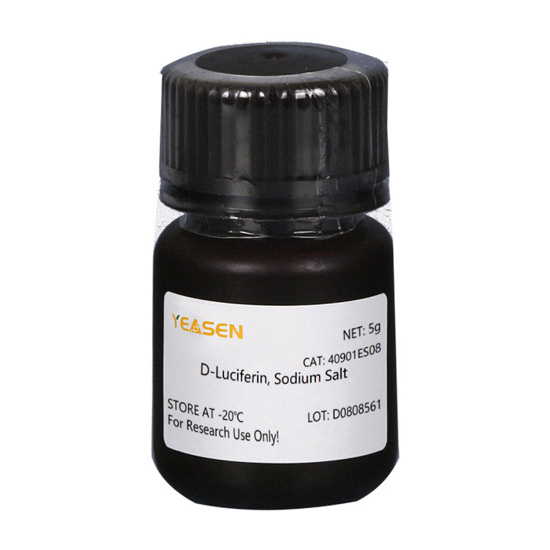
D-luciferin is a common substrate for Luciferase and is widely used throughout biotechnology, especially in vivo imaging technology. The mechanism of action is that luciferin (the substrate) is oxidized to emit light in response to ATP and luciferase (see figure below). It's produced when there's too much luciferin The light quantum number was positively correlated with the concentration of luciferase. Plasmids carrying luciferase encoding gene (Luc) were transfected into cells and introduced into study animals such as rats and mice In vivo, fluorescein is then injected and changes in light intensity are detected using bioluminescence imaging (BLI) to monitor disease progression or drug efficacy in real time and so on. ATP's influence on the reaction system can also be used to indicate energy or vital signs according to changes in bioluminescence intensity.

D-luciferin is also commonly used in in vitro studies, including luciferase and ATP levels analysis; Reporter gene analysis; High-throughput sequencing and various contamination tests. There are currently three product forms: D-luciferin (free acid), D-luciferin salts (sodium and potassium salts). The main difference lies in solubility: the former is water-soluble and the solubility of buffer system is weak, except soluble in weak bases such as low concentration NaOH and KOH solutions, soluble in methanol and DMSO; The latter is easily soluble in water or buffer, easy to use, solvent non-toxic, especially suitable for in vivo experiments. As a solution, there is no substantial difference between the three products in the vast majority of applications.
Features
High Purity: >99%
High Solubility: Up to 60 mg/mL in water
High Sensitivity: Capable of detecting as few as hundreds of cells; effective imaging depth of 3–4 cm
Stable Signal: High bioluminescence intensity and slow signal decay, ideal for large-scale in vivo validation studies
Application
In the tumorigenesis experiment in nude mice, the tumor growth was observed without invasion in real time, without tumor stripping measurement.
To test the effect of the administration on tumor growth or metastasis, the fluorescein substrate can be completely eliminated within 3 hours, without interference to the drug experiment.
The localization and distribution of foreign cells in animals were detected.
The target gene or promoter of the target gene is fused to the luciferase gene to detect changes in gene expression during drug treatment or disease progression.
Monitoring stem cell transplantation, survival and proliferation; Trace the distribution and migration of stem cells in vivo.
Specifications
| English synonym | (S)-4,5-Dihydro-2-(6-hydroxy-2-benzothiazolyl)-4-thiazolecarboxylic acid sodium salt; D-Luciferin firefly, sodium salt monohydrate; |
| CAS NO. | 103404-75-7 |
| Formula | NaC11H7N2O3S2·H2O |
| Molecular weight | 320.32 g/mol |
| Appearance | Light yellow powder |
| Solubility | Solube in water(100 mg/mL) |
| Purity (HPLC) | ≥95% |
Components
| Components No. | Name | 40901ES01 | 40901ES02 | 40901ES03 | 40901ES08 | 40901ES10 |
| 40901 | D-Luciferin, Sodium Salt | 100mg | 500 mg | 1g | 5g | 10g |
Figures
1. Superior Performance
(A). Superior Signal Stability

Figure 1. Superior Signal Stability Over Time
Fluorescence signal was measured every 5 min. Results showed that the signal from the Yeasen product decayed more slowly, with only about 20% reduction
(B). Higher Fluorescence Intensity

Figure 2. Higher Fluorescence Intensity
Fluorescence intensity test using a multifunctional microplate reader demonstrated that the Yeasen luciferin potassium salt produced stronger fluorescence signals compareafter 40 minutes.
2. Application Case
Cas 1. D-Luciferin is applicable in cancer research.

Figure 3. Mechanistic Study of Circular RNA in Tumor Growth.
Using Yeasen products, Gu Y, Wang Y, et al. investigated the role of circRNA in tumor progression. The study revealed that circular RNA circIPO11 drives self-renewal of liver cancer–initiating cells via Hedgehog signaling (Mol Cancer, 2021, 20(1):132).
Cas 2. D-Luciferin enables real-time monitoring of tumor growth.

Figure 4. In Vivo Pharmacodynamic Evaluation of Antitumor Drugs.
Yeasen products were applied in the study by Huang K, Liang Q, et al., which identified a novel allosteric inhibitor of phosphoglycerate mutase 1 (PGAM1) that suppresses growth and metastasis of non–small-cell lung cancer (Cell Metab, 2019, 30(6):1107–1119.e8).
Storage
FAQ
- Thiazoline-4-carboxylic acid.
1) Solubility: The salt form is easily soluble in water. The solubility of potassium salt is 60mg/ml, and that of sodium salt is 100mg/ml. Free acid is not easily soluble in water. It can be dissolved by a weak alkaline solution of sodium bicarbonate, and its solubility in methanol is 10mg/ml, and in DMSO is 50mg/ml.
2) Toxicity: The salt form is more convenient to use during the process, especially in in vivo imaging experiments, as it can dissolve in water and thus has less reaction toxicity (Fluorescein is a low-molecular-weight organic compound composed of phenylthiazole and thiazole carboxylic acid groups, which has low toxicity). 3) Usage effect: There is no significant difference. In in vivo experimental studies, the usage rate of potassium salt is higher.
1. Instruments with a biochemiluminescence detection module. The light produced by luciferin can be detected by a photometer or a scintillation counter. Common in vivo imaging instruments: such as the IVIS® Lumina small animal in vivo imaging system, and the In-Vivo Xtreme multi-mode small animal in vivo imaging instrument from Bruker, Germany.
2. Multi-functional microplate readers: It is necessary to confirm with the instrument manufacturer whether it is a specific biochemiluminescence detection module (note: cannot use a fluorescence microscope).
1) Injection method: It can be done through intraperitoneal injection or tail vein injection.
2) Injection volume: The scientific approach is to evaluate the injection dose based on the kinetic curve. The initial attempted injection dose was set at 150mg substrate/kg of mouse body weight. Therefore, the purchase quantity can be calculated according to the above method: If there are 10 mice, 22 - 25g, then 33 - 37.5mg of substrate is required.
Citations
Documents
| Brochure |
|---|
| In_vivo_imaging_Leaving_Tumor_Cells_Nowhere_to_Hide.pdf |


You may also like
Inquiry
FAQ
The product is for research purposes only and is not intended for therapeutic or diagnostic use in humans or animals. Products and content are protected by patents, trademarks, and copyrights owned by Yeasen Biotechnology. Trademark symbols indicate the country of origin, not necessarily registration in all regions.
Certain applications may require additional third-party intellectual property rights.
Yeasen is dedicated to ethical science, believing our research should address critical questions while ensuring safety and ethical standards.